Category Archives: Abdomen

How To Evaluate A Stab To The Diaphragm – Part 2

Yesterday I gave a little perspective on the use of CT in assessing the diaphragm after penetrating injury. Today, I’ll break it down into some practical steps you can follow the next time you see one.

Step 1. Stable or unstable? If your patient arrives with unstable vital signs, and there is no other source but the abdomen, the answer is simple. Go to the OR for a laparotomy. Period. They are exsanguinating and the hemorrhage needs to be stopped.

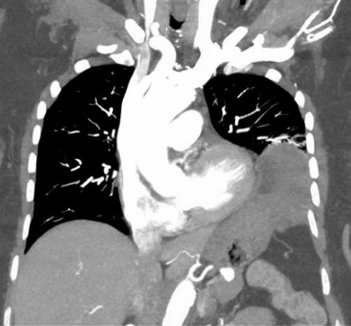
Step 2. Mark the sites of penetration and take a chest x-ray. This will let you evaluate the potential trajectory of the object, and will give you your first glimpse of the diaphragm.

Step 3. Examine the abdomen. Actually, you should be doing this at the same time you are setting up for Step 2. If your patient has peritoneal signs, no further evaluation is needed. Just go to the OR for laparotomy. Look at the chest x-ray once you get there.

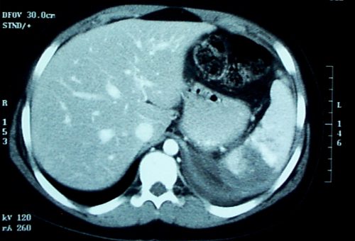
Step 4. Right side? If your appreciation of the path of penetration involves just the liver, take the patient to CT for evaluation of chest, abdomen, and pelvis. You need to see all three of these areas to assess for blood and fluid in both body cavities. After the study, if you still think the injury is limited to the liver, admit the patient for observation.

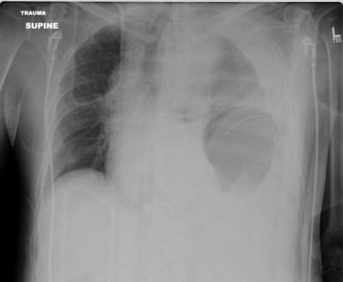
Step 5. Left side? Look at that chest x-ray again. If there are any irregularities at all, strongly consider going to the OR and starting with diagnostic laparoscopy. These irregularities can be glaring, like in the x-ray above. But they can be subtle, like some haziness above the diaphragm or small hemothorax. Obviously, if the injury is as clear as on the x-ray above, just open the abdomen. But if in doubt, start small. And remember my advice on “lunchothorax.”

Step 6. Admit and observe. Check the abdomen periodically, and repeat the chest x-ray daily. If anything changes, consider diagnostic laparoscopy. As a general rule, I don’t keep patients NPO “just in case.” Most will pass this test, and I don’t see a reason to starve my patients for the low likelihood they need to go to the OR.

Step 7. Make sure your patient gets a follow up evaluation. See them in your outpatient clinic, get a final chest x-ray and abdominal exam before you completely clear them.

Print Friendly, PDF & Email

How To Evaluate A Stab To The Diaphragm – Part 1

Penetrating injury to the diaphragm, and specifically stab wounds, have been notoriously hard to diagnose since just about forever. Way back in the day (before CT), we tried all kinds of interesting things to help figure out if the patient had a real injury. Of course, we could just go to the OR and lap the patient (laparoscopy did not exist then). But the negative lap rate was significant, so we tried a host of less invasive techniques.

Remember diagnostic peritoneal lavage? Yeah, we tried that. The problem was that the threshold for red cells per cubic mm was not well defined. Some would supplement this technique with a chest tube to see if lavage fluid would drain out. And one paper described instilling nuclear medicine tracer into the abdomen and sitting the patient under a gamma camera for a few hours to see if any ended up in the chest. Groan!

We thought that CT would save us. Unfortunately, resolution was terrible in the early years. If you could actually see the injury on CT, it was probably because a large piece of stomach or colon had already fallen through it. But as detectors multiplied and resolution improved, we could begin to see some smaller defects. But we still missed a few. And the problem is that left-sided diaphragmatic holes slowly enlarge over time (years), until the stomach or colon falls through it. (See below)

A group of radiologists and surgeons in a Turkish trauma hospital recently published a modest series of patients with left-sided diaphragm injuries evaluated by CT. They looked at about 5 years of their experience in a group of patient who were at risk for the injury due to a thoraco-abdominal stab wound. Unstable patients were immediately taken to OR. All of the remaining patients underwent an initial CT scan, followed by diagnostic laparoscopy after 48 hours if they remained symptom free.

Here are the factoids:

  • A total of 43 stable patients with a left thoraco-abdominal stab were evaluated
  • 30 patients had a normal CT, and 13 had the appearance of an injury
  • Of those who were CT positive, only 9 of 13 (69%) actually had the injury at operation
  • Two of the 30 (7%) who were CT negative were found to have a diaphragm injury during followup laparoscopy
  • So in the author’s hands, there was 82% sensitivity, 88% specificity, a positive predictive value of only  69%, and a negative predictive value of 93%

Bottom line: The authors somehow looked at the numbers and concluded that CT is valuable for detecting left diaphragm injury. Huh? They missed 7% of injuries, only finding them later at laparoscopy. And they had a 31% negative laparotomy rate. 

Now, it could be that the authors were using crappy equipment. Nowhere in their paper do they state how many detectors, or what technique was used. Since it took place over a 5 year period, it is quite possible that the earlier years of the study used equipment now considered to be out of date, or that there was no standardized technique.

CT may not yet be ready for prime time. But it can be a valuable tool. Tune in tomorrow for some tips on how and when to look for this insidious injury.

Reference: Evaluation of diaphragm in penetrating left thoracoabdominal
stab injuries: The role of multislice computed tomography. Injury 46:1734-1737, 2015.

Print Friendly, PDF & Email

Splenic Vascular Blush

Contrast blush is always a concern when seen on CT of the abdomen for trauma. It can represent one of two things, and both are bad:

  • Active extravasation of contrast
  • Splenic pseudoaneurysm

These two clinical issues can be distinguished by looking at the location of the contrast and its persistence. A pseudoaneurysm is located within the parenchyma, and the contrast will wash away, so it will not be visible on delayed images. Contrast that extends beyond the parenchyma or persists in delayed views represents active bleeding. In either case, the failure rate of nonoperative management exceeds 80% in adults without additional measures being taken.

Clinically, these patients usually act as if they are losing volume and require additional crystalloid and/or blood transfusion. The natural history in adults is for bleeding to continue or for the pseudoaneurysm to rupture, resulting in a quick trip to the operating room.

If vital signs can be maintained with fluids and blood, a trip to interventional radiology may solve the problem. Selective or nonselective embolization can be carried out and patients with only a few bleeding points can be spared operation. However, if multiple bleeding areas are seen, it is probably better to head to the OR for splenorrhaphy or splenectomy.

The image below shows likely areas of extravasation. They are a bit large to be pseudoaneurysms.

Spleen Blush-CT

Children are different than adults. Extravasation from spleen injuries in prepubescent children frequently stops on its own. Angiography should only be used if the child is failing nonoperative management.

Next post: A new paper looks at the natural history of these lesions.

Print Friendly, PDF & Email

Could Be A Urethral Injury, But The Catheter’s Already In?

You’re seeing a trauma patient, probably a transfer from somewhere else. Either they told you there “may have been” some blood at the tip of the urethra, or maybe you see it smearing the outside of a urinary catheter that’s already in place! How do you proceed from here?

First, try not to get into that situation. Make sure that everyone on your team knows that gross blood at the meatus, male or female, means urethral injury until proven otherwise. If it’s not gross blood, it could be that the patient was incontinent and has hematuria from other causes. The fear with passing a catheter across a urethral injury is that it may convert a partial tear to a complete one. Reconstruction and complications from the latter are far more serious.

But the catheter is there. What to do?

First, leave the catheter in place. You must assume that the injury is present, and you need to rule it in or rule it out in order to decide what to do with the catheter. If the injury is not really there, then you can remove the catheter when indicated. If it really is present, then the urethral injury is being treated appropriately.

Next, do a urethrogram. I’ve previously described how to do it here, but the technique I describe is only appropriate for uncatheterized patients. The technique must be modified to use thin contrast and a method to inject alongside the catheter. To do this, fill a 20-30cc syringe with contrast (Ultravist or similar liquid) and put an 18 gauge IV catheter on the tip (no needles, please). Slide the IV catheter alongside the urinary catheter, clamp the meatus with your fingers, pull the penis to the side and inject under fluoroscopy. The contrast column will not be as vivid as with a regular urethrogram because it is outlining the urinary catheter, so there is less volume.

If the contrast travels the length of the urethra and enters the bladder without leaking out into soft tissue, there is no injury. If there is contrast leakage, stop injecting and plan to call a urologist.

Finally, be on the lookout for associated injuries. Urethral injuries are frequently found in patients with anterior pelvic fractures and perineal injuries.

Related post:

Link: blood at the urethral meatus (Atlas-Emerg-Medicine.org.ua from McGraw-Hill)
Print Friendly, PDF & Email

Thoughts On Traumatic Hematuria: Part 2

Yesterday, I discussed blood in the urine from a urethra. As I mentioned, there is typically not much from that particular injury. Today, I’ll dig into the three causes of real hematuria.

All of these tubes show gross hematuria except the one on the right.

  • Bladder injury. This can occur with either blunt or penetrating injury. The degree of hematuria is variable with stabs or gunshots, but tends to be much darker in blunt injury. This happens because the size of the bladder injury tends to be greater with blunt force. The bladder injury is not necessarily full-thickness with blunt trauma. It may just be some wall contusion and underlying mucosal injury. But frequently, with seat belt injury and/or A-P compression injuries to the pelvis (“open book”), the injury is full thickness.
    • Tip: If less than 50cc of very dark urine flow from the catheter upon insertion, it is likely that your patient has an intraperitoneal bladder rupture!
  • Ureteral injury. This injury is very rare. The most common mechanism is penetrating, but this structure is so small and deep that it seldom gets hit by naything. Patients with multiple lumbar transverse process fractures will occasionally have a small amount of hematuria, probably from a minor contusion. More often than not, the hematuria is microscopic, so we should never know about it.
  • Kidney injury. The most important fact regarding renal injury is that the degree of injury has no correlation with the amount of hematuria. The most devastating injury, a devascularized kidney, frequently has little if any gross hematuria. And conversely, a very minor contusion can produce very red urine.

So what about diagnosis? It’s easy! If you see gross hematuria, insert a foley catheter (if not already done) and order a CT of the abdomen/pelvis with contrast, as well as a CT cystogram. The latter must not be done using passive filling of the bladder with a clamped catheter. Contrast must be infused into the bladder under pressure to ensure a bladder injury can be identified.

CT scan is an excellent tool for defining injuries to kidney, ureter, and bladder, and will identify extravasation into specific places and allow grading. Specific management will be the topic of future posts.

Print Friendly, PDF & Email