All posts by The Trauma Pro

How Does It Work? The Lowly Blood Pressure Cuff

The blood pressure cuff is one of those devices trauma professionals don’t give a second thought to. Old timers like me remember using the cuff with a sphygmomanometer and stethoscope to get manual blood pressures. I’ve had to do this twice in recent months on airplanes, and I had forgotten how much work this is.

But technology makes things easier for us. Now you just slap a cuff on the arm (or wrist), push a button, and voila! You’ve got the pressure.

But have you stopped to think about how this actually works? Why don’t we need the stethoscope any more? Here’s the scoop:

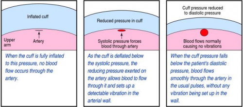
When you take a manual blood pressure, the cuff is inflated until a pulse can no longer be auscultated with the stethoscope. The pressure is slowly released using a little thumb wheel while listening for the pulse again. The pressure at which it is first audible is the systolic, and the pressure at which it softens and fades away is the diastolic.

The automatic blood pressure device consists of a cuff, tubing that connects it to the monitor, a pressure transducer in line with the tubing, a mini air pump, and a small computer. The transducer replaces the analog pressure gauge, and the pump and computer replace the human.

The transducer can “see” through the tubing and into the cuff. It is very sensitive to pressure and pressure changes. The computer directs the pump to inflate to about 20 torr above the point where pulsations in the air column cease. It then releases the pressure at about 4 torr per second, “feeling” for air column vibrations to start. When this occurs, the systolic pressure is recorded. Deflation continues until the vibrations stop, representing the diastolic pressure.

bpcuff

Piece of cake! But here’s the question: is it accurate? Tomorrow, I’ll write about how the automated cuff compares to an indwelling arterial line.

Related post:

How Many Salt Tabs In A Liter Of Saline?

Seems like a simple, silly question, right? I dare you to figure it out without reading this post!

horse-salt-block-lick2

On occasion, our brain injured trauma patients have sodium issues. You know, cerebral salt wasting. Trying to maintain or regain the normal range, without making any sudden moves can be challenging. There are a lot of tools available to the trauma professional, including:

  • Saline
  • Hypertonic saline
  • Salt tablets
  • Fluid restriction
  • Some combination thereof

Fun times are had trying to figure out how much extra sodium we are giving with any of the first three items. This is important as you begin to transition from the big guns (hypertonic), to regular saline, and then to oral salt tabs.

Below is a quick and dirty conversion list. I won’t make your heads explode by trying to explain the math involved changing between meq, mg, moles, sodium and sodium chloride.

  • The “normal saline” bags we use are actually 0.9% saline (9 gm NaCl per liter)
  • Hypertonic saline can be 3% or 5% (30 gm or 50 gm per liter)
  • Salt tabs are usually 1 gm each (and oh so yummy)

Therefore, a liter of 0.9% normal saline is the same as 9 salt tabs.

A liter of 3% hypertonic saline is the same as 30 salt tabs. The usual 500cc bag contains 15.

A liter of  5% hypertonic saline is the same as 50 salt tabs. The usual 500cc bag contains 30.

To figure out how many tablets you need to give to match their IV input, calculate the number of liters infused, then do the math! And have fun!

Next Trauma MedEd Newsletter Is Coming Next Week!

As promised, the next Trauma MedEd newsletter will be released next week. Just in time for some light Christmas reading!

The topic is “Prevention.” Here are the areas I’ll be covering:

  • The American College of Surgeons requires all US trauma centers to engage in prevention activities. Unfortunately, there is frequently confusion about the role of the injury prevention coordinator, what kinds of programs are acceptable, and how local data needs to be included in prevention planning. I will cover all of this, and more, in the first part of the newsletter.
  • Curious about what others are doing out there? I’ll give you an idea of the most common prevention programs, and whether they are national programs or home grown.
  • I’ll review a few papers on the efficacy of trauma prevention programs.
  • Finally, I’ll give some tips on how to optimize the performance of your injury prevention coordinator and design effective programs.

As always, this issue will go to all of my subscribers first. If you are not yet one of them, click this link to sign up and/or download back issues.

Unfortunately, non-subscribers will have to wait until I release the issue on this blog, sometime during the week after Christmas. So sign up now!

The IVC Filter In Trauma: Why?

The inferior vena cava (IVC) filter has been around in one form or another for over 40 years. One would think that we would have figured everything about it out by now. But no!  The filter has evolved through a number of iterations and form factors over the years. The existing studies, in general, give us piecemeal information on the utility and safety of the device.

One of the major innovations with this technology came with the development of a removable filter. Take a look at the product below. Note the hook at the top and the (relatively) blunt tips of the feet. This allows a metal sheath to be slipped over the filter while in place in the IVC. The legs collapse, and the entire thing can be removed via the internal jugular vein.

ivc-filter-complications1

The availability of the removable filter led the American College of Chest Physicians to recommend their placement in patients with known pulmonary embolism (PE) or proximal deep venous thrombosis (DVT) in patients with contraindications to anticoagulation. Unfortunately, this has been generalized by some trauma professionals over the years to include any trauma patients at high risk for DVT or PE, but who don’t actually have them yet.

One would think that, given the appearance of one of these filters, they would be protective and clots would get caught up in the legs and be unable to travel to the lungs as a PE. Previous studies have taught us that this is not necessarily the case. Plus, the filter can’t stop clots that originate in the upper extremities from becoming an embolism. And there are quite a few papers that have demonstrated the short- and long-term complications, including clot at and below the filter as well as post-phlebitic syndrome in the lower extremities.

A new study from Boston University reviewed their own experience retrospectively over a 9 year period. This cohort study looked at patients with and without filters, matching them for age, sex, race, and injury severity. The authors specifically looked at mortality, and used four study periods during the 9 year interval.

Here are the factoids:

  • Over 18,000 patients were admitted during the study period, resulting in 451 with an IVC filter inserted and 1343 matched controls
  • The patients were followed for an average of 4 years after hospitalization
  • Mortality was identical between patients with filters vs the matched controls

dvt-study

  • There was still no difference in mortality, even if the patients with the filter had DVT or PE present when it was inserted
  • Only 8% ever had their “removable” filter removed (!)

Bottom line: Hopefully, it’s becoming obvious to all that the era of the IVC filter has come and gone. There are many studies that show the downside of placement. And there are several (including this one) that show how forgetful we are about taking them out when no longer needed. And, of course, they are expensive. But the final straw is that they do not seem to protect our patients like we thought (hoped?) they would. It’s time to reconsider those DVT/PE protocols and think really hard about whether we should be inserting IVC filters in trauma patients at all.

Related post:

Reference: Association Between Inferior Vena Cava Filter Insertion
in Trauma Patients and In-Hospital and Overall Mortality. JAMA Surg, online ahead of print, September 28, 2016.

(Mis)Use of Helicopter Transport For Pediatric Trauma

Helicopter transport is an integral and important part of modern day trauma care. Since the inception helicopter emergency medical services (HEMS) for civilian use in the 1970’s, its use has been steadily increasing. And it’s expensive, at least five times more costly than ground transport. Plus, there are risks to both crew and patient, in that there have been 200 deaths of both patients and flight crews. Indeed, flight crews have one of the riskiest jobs, with 5 times more on-the-job deaths than police officers.

So it becomes very important to make sure that this mode of transport is justified. As I wrote previously, the adult HEMS literature is extensive, but not terribly convincing. There is far less data available regarding pediatric patients. And the data that does exist suggests that there may be significant overtriage and overuse.

A study using the National Trauma Data Bank (NTDB) was performed by researchers at Duke University. They reviewed the data for a 5 year period (2007-2011), which is fairly old in my opinion. And they included “children” up through age 18, which are also a bit old, in my opinion. Since there are no real quantitative criteria for overtriage in place, the authors picked three: low injury severity (ISS<10), normal physiology (RTS=12), and low predicted mortality using TRISS (<5%). A total of 127,489 patient records were analyzed.

Here are the factoids:

  • 14% arrived via helicopter EMS,  56% by ground EMS, and 29% by private vehicle or walk-in
  • HEMS patients were more likely to have head, thoracic, or abdominal injuries, and overall severe injuries (good!)
  • Adjusted mortality for patients transported by air was significantly less than for ground (really good)
  • 38% of HEMS patients had ISS < 9, and 66% had completely normal physiology (bad)
  • Overall, 32% to 82% of children did not meet criteria for appropriate transport

Bottom line: There are a number of flaws in this study that could be improved upon. However, it does provide some interesting data. Helicopter transport does save lives in the younger population, and was estimated at 2 per 100 flights. This is very promising. However, offsetting this was the fact that nearly half of transports failed one or more arbitrary appropriateness criteria. The recommendations I published yesterday need to be adopted, and both state trauma systems and local EMS agencies need to develop and enforce guidelines to optimally use this valuable and expensive resource.

Reference: Current use and outcomes of helicopter transport in pediatric trauma: a review of 18,291 transports. J Ped Surg in press 27 Oct 2016.