All posts by The Trauma Pro

Serial Hemoglobin / Hematocrit – Huh? Part 2

In my last post, I waxed theoretical. I discussed the potential reasons for measuring serial hemoglobin or hematocrit levels, the limitations due to the rate of change of the values, and conjectured about how often they really should be drawn.

And now, how about something more practical? How about an some actual research? One of the more common situations for ordering serial hemoglobin draws occurs in managing solid organ injury. The vast majority of the practice guidelines I’ve seen call for repeating blood draws about every six hours. The trauma group at the University of Florida in Jacksonville decided to review their experience in patients with liver and spleen injuries. Their hypothesis was that hemodynamic changes would more likely change management than would lab value changes.

They performed a retrospective review of their experience with these patients over a one year period. Patients with higher grade solid organ injury (Grades III, IV, V), either isolated or in combination with other trauma, were included. Patients on anticoagulants or anti-platelet agents, as well as those who were hemodynamically unstable and were immediately operated on, were excluded.

Here are the factoids:

  • A total of 138 patients were included, and were separated into a group who required an urgent or unplanned intervention (35), and a group who did not (103)
  • The intervention group had a higher ISS (27 vs 22), and their solid organ injury was about 1.5 grades higher
  • Initial Hgb levels were the same for the two groups (13 for intervention group vs 12)
  • The number of blood draws was the same for the two groups (10 vs 9), as was the mean decrease in Hgb (3.7 vs 3.5 gm/dl)
  • Only the grade of spleen laceration predicted the need for an urgent procedure, not the decrease in Hgb

Bottom line: This is an elegant little study that examined the utility of serial hemoglobin draws on determining more aggressive interventions in solid organ injury patients. First, recognize that this is a single-institution, retrospective study. This just makes it a bit harder to get good results. But the authors took the time to do a power analysis, to ensure enough patients were enrolled so they could detect a 20% difference in their outcomes (intervention vs no intervention). 

Basically, they found that everyone’s Hgb started out about the same and drifted downwards to the same degree. But the group that required intervention was defined by the severity of the solid organ injury, not by any change in Hgb.

I’ve been preaching this concept for more than 20 years. I remember hovering over a patient with a high-grade spleen injury in whom I had just sent off the requisite q6 hour Hgb as he became hemodynamically unstable. Once I finished the laparotomy, I had a chance to pull up that result: 11gm/dl! 

Humans bleed whole blood. It takes a finite amount of time to pull fluid out of the interstitium to “refill the tank” and dilute out the Hgb value. For this reason, hemodynamics will always trump hemoglobin levels for making decisions regarding further intervention. So why get them?

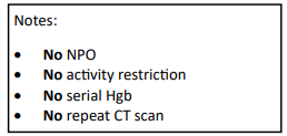
Have a look at the Regions Hospital solid organ injury protocol using the link below. It has not included serial hemoglobin levels for 18 years, which was when it was written. Take care to look at the little NO box on the left side of the page.

I’d love to hear from any of you who have also abandoned this little remnant of the past. Unfortunately, I think you are in the minority!

Reference: Serial hemoglobin monitoring in adult patients with blunt solid organ injury: less is more. J Trauma Acute Care Open 5:3000446, 2020.

Serial Hemoglobin / Hematocrit – Huh? Part 1

The serial hemoglobin (Hgb) determination. We’ve all done them. Not only trauma professionals, but other in-hospital clinical services as well. But my considered opinion is that they are not of much use. They inflict pain. They wake patients up at inconvenient hours. And they are difficult to interpret. So why do them?

I’m reposting this mini-series on serial hemoglobin draws in light of a new paper that was published in the Journal of Trauma Nursing. It continues next week with Part 2, and then a summary of the new paper.

First, what’s the purpose? Are you looking for trends, or for absolute values? In trauma, the most common reason to order is “to monitor for bleeding from that spleen laceration” or some other organ or fracture complex. But is there some absolute number that should trigger an alarm? If so, what is it? The short answer is, there is no such number. Patients start out at a wide range of baseline values, so it’s impossible to know how much blood they’ve lost using an absolute value. And we don’t use a hemoglobin or hematocrit as a failure criterion for solid organ injury anymore, anyway.

What about trends, then? First, you have to understand the usual equilibration curve of Hgb/Hct after acute blood loss. It’s a hyperbolic curve that reaches equilibrium after about 3 days. So even if your patient bled significantly and stopped immediately, their Hgb will drop for the next 72 hours anyway. If you really want to confuse yourself, give a few liters of crystalloid on top of it all. The equilibration curve will become completely uninterpretable!

And how often should these labs be drawn? Every 6 hours (common)? Every 4 hours (still common)? Every 2 hours (extreme)? Draw them frequently enough, and you can guarantee eventual anemia.

Bottom line: Serial hemoglobin/hematocrit determinations are nearly worthless. They cost a lot of money, they disrupt needed rest, and no one really knows what they mean. For that reason, my center does not even make them a part of our solid organ injury protocol. If bleeding is ongoing and significant, we will finding it by looking at vital signs and good old physical exam first. But if you must, be sure to explicitly state what you will do differently at a certain value or trend line. If you can’t do this and stick to it, then you shouldn’t be ordering these tests in the first place!

In my next post, I’ll discuss a paper that objectively shows the (lack of) utility of this testing method.

Creating A Virtual RTTDC Course

The Rural Trauma Team Development Course (RTTDC) was introduced by the American College of Surgeons (ACS) to improve the care of trauma patients in rural communities. It is a staple of education for Level III and IV trauma centers in rural areas. Like everything else, most courses were shut down by the COVID-19 pandemic.

Conemaugh Memorial Medical Center in Johnstown, Pennsylvania, polled its local referral hospitals and discovered that the majority felt a significant need for continuing, in-person education that was not being met. This need, coupled with the observation of an increased number of opportunities for improvement in patients transferred to them, led them to consider adapting the RTTDC to a virtual format so the course could continue.

Since RTTDC is a product of the ACS, it is no simple matter to change it in any way. The trauma program worked with the ACS to get permission to make changes to the course.  Speakers with specialization in their topic recorded all of the lectures. They contained embedded questions to be answered using the polling feature of the Zoom software used.

The most challenging adaptation was simulation development for the hands-on portions of the course. These were painstakingly recorded on video in a simulation laboratory and incorporated into the lecture material.

Preregistration was brisk, and 41 participants signed up for the course. The format consisted of a lecture with live discussion and participant questions, followed by a simulation video moderated by the course director. All questions were answered before moving on to the next module.

Several positive changes were noted in the months following the course:

  • Many facilities purchased additional equipment, such as traction splints, pelvic binders, and blood warmers.
  • Some hospitals began acquiring tranexamic acid and prothrombin concentrate.
  • One facility modified its radiographic imaging policy.
  • All hospitals tightened their performance improvement processes and began to identify more opportunities for improvement.

Of course, some downsides were also identified:

  • Production of the course was very intensive and administratively challenging.
  • There was the possibility of teleconferencing hardware/software failure.
  • It was difficult for the presenters to “read the audience” because of the Zoom headshot.
  • Truly interactive discussions were difficult to achieve.

Bottom line: This is a creative example of a rural trauma center identifying regional needs and developing an innovative solution despite the pandemic. Despite the amount of work needed to pull it off, the results were very positive. Although the course should ideally be produced in person, this may not be feasible in some very remote areas. 

Hopefully, the ACS will be able to recognize this work and the need for this format. It should create a virtual version to help spread the word to all rural trauma centers.

Reference: Virtual Rural Trauma Team Development Course: Trying To Zoom In On A Solution. J Trauma Nursing 20(3):186-190, 2023.

Leukocytosis After Splenic Injury

Any trauma professional who has dealt with spleen injuries knows that the white blood cell (WBC) count rises afterwards. And unfortunately, this elevation can be confusing if the patient is at risk for developing inflammatory or infectious processes that might be monitored using the WBC count.

Is there any rhyme or reason to how high WBCs will rise after injury? What about after splenectomy or IR embolization? An abstract is being presented at the Clinical Congress of the American College of Surgeons next month that examines this phenomenon.

This retrospective study looked at a convenience sample of 75 patients, distributed between patients who had splenic injury that was either not treated, removed (splenectomy), or embolized. Data points were accumulated over 45 days.

Here are the factoids:

  • 20 patients underwent splenectomy, 22 were embolized, and 33 were observed and not otherwise treated
  • Injury severity score was essentially identical in all groups (19)
  • Splenectomy caused the highest WBC counts at the 30 day mark (17.4K)
  • Embolized patients had mildly elevated WBC levels (13.1K) that were just above the normal range at 30 days
  • Observed patients had high normal WBC values (11.0K) after 30 days
  • Values in observed and embolized patients normalized to about 7K after 30 days; splenectomy patient WBC count remained mildly elevated at 14.1K.
  • The authors concluded that embolization does not result in permanent loss of splenic function (bad conclusion, rookie mistake!)

Bottom line: This study is interesting because it gives us a glimpse of the time course of leukocytosis in patients with injured spleens. If you need to follow the WBC for other reasons, if gives a little insight into what might be attributable to the spleen. Splenectomy generally results in a chronically elevated WBC count, which tends to vary in the mid-teens range. Embolization (in this study) transiently elevates the WBC count, but it then drops back to normal.

The big problem with this study (besides it being small) is that it fails to recognize that there are many different shades of embolization. Splenic artery? Superselective? Selective? I suspect that the WBC count in main splenic artery embolization may behave much like splenectomy in terms of leukocytosis. And the conclusion about splenic function being related to WBC count was pulled out of a hat. Don’t believe it.

Reference: Leukocytosis after Splenic Injury: A Comparison of Splenectomy, Embolization, and Observation. American College of Surgeons Scientific Forum Abstracts pg S164, 2015.

How Fast Do Trauma Patients Die?

For years, I’ve taught my residents participating in trauma activations, “Your patient is bleeding to death until proven otherwise.” This concept served as the basis of the [poorly documented] “Golden Hour” and for decades has directed our efforts at getting patients to a center with an immediately available OR as quickly as possible.

Donald Trunkey published the first paper illustrating the trimodal distribution of death in 1983 in Scientific American. A crude graph showed the large spike in early deaths that occurred within this first hour. But the paper was mainly observational and was not based on quantitative data.

Wouldn’t it be nice to know how quickly these injured patients were dying, and of what? The trauma group at the University of Pennsylvania massaged data in the state trauma database, focusing on patients who died of their injuries during the first four hours. They created two variables to more objectively compare times, the TD5 and the TD50. These are the time at which 5% and the time at which 50% (median) had died, respectively.

The Pennsylvania Trauma Outcomes Study database contains a huge amount of data. During the 11 years of the study, a total of 6,547 met the mortality criteria for analysis.

Here are the factoids:

  • The mechanism of injury was about 60% blunt / 40% penetrating, with an average ISS of 33
  • The majority of these patients (85%) were hypotensive before their death, meaning that they were likely bleeding to death on arrival
  • The  overall TD5 was 23 minutes, and the TD50 was 59 minutes
  • These numbers were shorter for penetrating injuries, TD5=19 minutes and TD50=43 minutes
  • Patients who were not hypotensive lived a little longer: TD5=44 minutes and TD50 = 2 hours 18 minutes
  • 77% of patients died in the ED and 19% in the OR. The remainder died in the ICU.

This chart shows the TD5 by mechanism and type of surgery. This represents when after arrival, patients start dying due to their injuries. Penetrating injury plus hypotension kills the fastest at 19 minutes and head injuries the slowest at 1:20.

Bottom line: The authors clearly show how soon seriously injured patients start to die. It’s less than 20 minutes in victims of penetrating injury with early hypotension. And the time between the “just start do die” point (TD5) and the “half are dead” point (TD50) is frighteningly short, just an additional twenty minutes!

There appears to be a bit of a grace period in patients who arrive with a normal blood pressure. Their TD50 is extended out to about two hours. All this means is that they are bleeding more slowly, but it is still killing them.

A good rule of thumb is that ANY hypotensive patient should make you justify why you are NOT ALREADY IN THE OPERATING ROOM! Dawdling in the trauma bay or performing unnecessary scans will push your patient much closer to the point of no return. Look at the huger percentage of patients in this study who died in the ED.

Remember, your patient is bleeding to death in front of your eyes, and the only place you can stop it is the OR!

Reference: Defining the optimal time to the operating room may
salvage early trauma deaths, J Trauma 76(5):1251-1258, 2014.