All posts by The Trauma Pro

Delayed Presentation Of Right Diaphragm Injury

Diaphragm injury from blunt trauma is uncommon, occurring in only a few percent of patients after high-energy mechanisms. They usually occur on the left side and are more frequently seen after t-bone type car crashes and in pedestrians struck by a car.

Blunt diaphragm injury on the right side is very unusual. Even so, it is more easily detected due to obvious displacement of the liver that can be seen on chest x-ray. Blunt injuries on the right side usually result in a large rent in the central tendon or detachment of the diaphragm from the chest wall. This allows the liver to herniate into the chest, and the chest x-ray finding is not subtle.
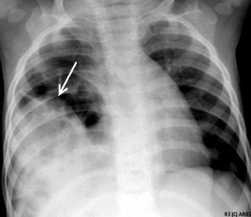
This image shows an acute herniation of the liver through the diaphragm. Due to the size of the liver, only part of it can typically fit through the rent. Radiologists call this the “cottage loaf” sign. Why? Here’s the bakery item it is named after. Get it now?

Thankfully, most of these injuries are identified in the acute setting. They must be addressed surgically because, if left untreated, more and more of the liver will slowly move into the chest resulting in respiratory problems in the long run.

Acute management usually consists of laparotomy to address both the diaphragm tear and any other associated intra-abdominal injuries. The liver should be reduced by sliding a hand next to it laterally into the chest cavity and pushing the dome downwards. The right triangular ligaments should be taken down (if they are not already destroyed) to mobilize the organ better so the diaphragm laceration can be closed. This is typically accomplished with some type of large (size 0) permanent suture. A chest tube will be needed to evacuate the iatrogenic pneumothorax created by opening the abdomen.

Chronic right diaphragm injuries are a different animal entirely. There is no longer any need to evaluate for intra-abdominal injury, so the procedure is usually performed through the chest. For smaller injuries, thoracoscopic procedures have been described that push the liver downwards and then either suture the diaphragm primarily or (more likely) incorporate a piece of mesh.

Larger injury requires conversion to an open procedure so more muscle power can be used to push the liver downwards to facilitate the repair. However, do not underestimate the adhesions that will be present between diaphragm and liver (and possibly the lung) in long-standing injuries. It may take some time to dissect them away. Rarely, a laparotomy (or laparoscopy) may be needed to assist for very large and complex injuries.

References:

  • Management of Delayed Presentation of a Right-Side Traumatic
    Diaphragmatic Rupture. World J Surg 36:260-265, 2012.
  • Delayed Discovery of Diaphragmatic Injury After Blunt Trauma:
    Report of Three Cases. Surg Today 35:407-410, 2005.

Guidelines For Diagnosis Of Diaphragmatic Injury

In today’s post, I will review the diaphragmatic injury practice guidelines published by the Eastern Association for the Surgery of Trauma (EAST).  I will follow this up on Friday with an interesting delayed diaphragm injury case.

Diaphragm injury is a troublesome one to diagnose. It is essentially an elliptical sheet of muscle that is doubly curved, so it does not lend itself well to diagnosis by axial imaging. The addition of sagittal and coronal reconstructions to a thoracoabdominal CT has been helpful but still has a far from perfect diagnostic record.

From an evaluation standpoint, there are several possibilities:

  • Observation – not generally recommended. It is usually combined with imaging such as a chest x-ray to see if interval changes occur that would indicate the injury.
  • Chest x-ray – this is not often diagnostic, but when herniation of abdominal contents is obvious, the patient most assuredly has an operative problem.
  • Thoracoabdominal CT scan – this technology keeps improving, especially with thinner cuts and different planes of reconstruction. Sometimes even subtle injuries can be detected. But this exam is still imperfect.
  • Laparoscopy or thoracoscopy – this technique yields excellent accuracy when the injury is in an area that can be viewed from the operative entry point chosen.
  • Laparotomy or thoracotomy – this is the ultimate choice and should be nearly 100% accurate. It is almost the most invasive and has more potential associated complications.

EAST reviewed a large body of literature and selected 56 pertinent papers for their quality and design. Then, they critically reviewed them and applied a standard methodology to answer several questions.

Here are the questions with the recommendations from EAST, along with my comments:

  • Should laparoscopy or CT be used to evaluate left-sided thoracoabdominal stab wounds? First, these patients must be hemodynamically stable and not have peritonitis. If either is present, there is no further need for diagnosis; a therapeutic procedure must be performed.
    Left-sided diaphragm injuries from stabs are evil. The hole is small, and since the pressure within the abdomen is greater than the chest, things always try to wiggle their way through this small hole. It can remain asymptomatic if the wiggler is just a piece of fat, but it can be catastrophic if a bit of the stomach or colon pushes through and becomes strangulated. Furthermore, these holes enlarge over time, so more and more stuff can push up into the chest.
    EAST recommends using laparoscopy for evaluation to decrease the incidence of missed injury. However, if the injury is in a less accessible location (posterior), the patient has body habitus issues or adhesions from previous surgery may lead to incomplete evaluation, laparotomy should be strongly considered.
  • Should operative or nonoperative management be used to evaluate right-sided thoracoabdominal penetrating wounds? Note that this is different than the last question. All penetrating injuries (stabs and gunshots) are included, and this one is for management, not evaluation. And the same caveats regarding hemodynamic stability and peritonitis apply. Again, it applies to both stabs and gunshots.
    Unlike left-sided injuries, right-sided ones are much more benign. This is because the liver keeps anything from pushing up through small holes, and they do not tend to enlarge over time due to this protection. For that reason, EAST recommends nonoperative management to reduce operation-related mortality and morbidity.
  • Should stable patients with acute diaphragm injury undergo repair via an abdominal or thoracic approach? This question applies to any diaphragm injury that requires an operation, such as a right-sided penetrating injury or any blunt injury. EAST recommends an approach from the abdomen to reduce morbidity and mortality. However, since abdominal injury frequently occurs in these cases, an approach from the chest limits the ability to identify and repair abdominal injuries. Otherwise, you may find yourself doing a laparotomy in addition to the thoracotomy.
  • Should patients with delayed visceral herniation through a diaphragm injury undergo repair via an abdominal or thoracic approach?  For years, the preferred approach for delayed presentations has been through the chest because the injury is easier to appreciate and repair.  However, if ischemic or gangrenous viscera are present, it will be more challenging to manage and repair from the chest. EAST does not make a specific recommendation for this question and suggests the surgical approach be determined on a case-by-case basis.
  • Should patients with an acute diaphragm injury from penetrating injury without concern for other intra-abdominal injuries undergo open or laparoscopic repair? The quality and quantity of data addressing this question were very low, but EAST recommends laparoscopy to repair these injuries to reduce morbidity and mortality. This includes blunt injuries, which tend to be larger. There were some conversions to an open procedure, especially in the blunt cases. The usual caveats on exposure, injury location, body habitus, and previous surgery apply.

Reference: Evaluation and management of traumatic diaphragmatic injuries: A Practice Management Guideline from the Eastern Association for the Surgery of Trauma. J Trauma 85(1):198-207.

Time Is Spine: Spinal Decompression For Central Cord Syndrome

Over the recent decades, there has been a huge push toward “evidence-based” medicine. Unfortunately, the available amount of high-quality literature is relatively low. And the field of neurotrauma is even less-represented than most.

Debates have raged over the years regarding the proper timing of surgical spine decompression in patients with spinal cord injuries. Many proponents of early decompression (within 24 hours) believe this may limit secondary injury to the cord. But there are also others who don’t buy into this idea.

Central cord syndrome is a special case of spinal cord injury. It is a partial injury, usually in the cervical spine. It causes varying degrees of pain, paresthesia, and paresis and usually affects the upper extremities more than the lowers.

Many neurosurgeons choose a “wait and see” attitude with central cord syndrome patients. However, a group of neurosurgeons spanning trauma hospitals in Canada, Philadelphia, and Baltimore published an interesting paper last year. They performed a propensity score-matched cohort study, comparing functional recovery in patients undergoing early vs. late decompression after sustaining a central cord injury.

They specifically selected patients from three national spinal cord injury databases who had a full ASIA impairment scale examination performed within 14 days of injury. Patients had to have AIS grade C or D, meaning that some motor function was still present, and had to show a major difference between upper and lower extremity motor strength.

Here are the factoids:

  • Of a combined dataset of 1692 patients, 300 met baseline criteria. However, only 186 were eligible for propensity score matching, with half in each study group (early vs. late decompression).
  • Follow-up data was only available in 148 patients, which was right at the limit of the authors’ power calculation
  • Early surgery was significantly associated with improved upper limb motor function recovery but not the lower extremity. Overall motor score was not improved.
  • There was no functional improvement after late surgery
  • Patients with higher ASIA score (D) showed no improvement, regardless of surgical timing
  • Patients with AIS C lesions had significant recovery of their motor score in both upper and lower extremities but not their FIM motor score
  • A higher percentage of early-surgery patients achieved complete independence, especially those involving upper extremity function. However, this did not reach statistical significance.

Bottom line: Spine decompression timing remains very controversial, with every neurosurgeon having their own opinion. Unfortunately, this study was borderline underpowered, which may have weakened its results. 

Several important trends were noted, however. First, early surgery did have an impact on functional recovery, especially in the upper extremities. This is especially important because use of the hands is critical to functional independence.  But the most exciting result was the trend toward a higher percentage of patients achieving complete independence after early surgery. To be clear, this was just a trend and did not achieve statistical significance.

It’s time to start working with our neurosurgery colleagues and nudging them toward considering earlier surgery on this subset of spinal cord injury patients. It will take time and education, but these patients will actually be able to thank us, especially if they are actually able to shake our hands!

Reference: Early vs Late Surgical Decompression for Central Cord Syndrome. JAMA Surg. 2022;157(11):1024-1032. doi:10.1001/jamasurg.2022.4454

The End Of Serial Hemoglobin/Hematocrit In Solid Organ Injury

Here’s the final post on my series covering serial hemoglobin testing in the management of solid organ injury.

We developed our first iteration of a solid organ injury practice guideline at Regions Hospital way back in 2002. It was borne out of the enormous degree of clinical variability I saw among my partners. We based it on what little was publicly available, including an EAST practice guideline.

Recognizing that the EAST guideline couldn’t dictate bedside care, we gathered together to meld it with our own clinical experience. We fashioned our first practice guideline later that year and tested it.  It included instructions for bedrest (only overnight), vital signs monitoring, and lab testing (on admission and once the next day).

That last bit about serial lab tests is an important one. We had seen anecdotal evidence in our patients that it wasn’t very helpful. For example, I had one patient in the ICU whose serial Hgb had just returned normal. However, a minute later they experienced a hard hypotensive episode, and I took him immediately to the OR and took out a ruptured and bleeding spleen.

I’ve written several posts on how quickly Hgb changes after hemorrhage. Unfortunately, this lab test just lags too long to be a reliable indicator of anything. A very recent study has been published by Texas Health Presbyterian in Dallas. The retrospectively reviewed patients with liver or spleen injury over five years. They examined how often serial hemoglobin determinations influenced management during the study period. Possible interventions were none, operation, angioembolization, or blood transfusion.

Here are the factoids:

  • There were 143 patients enrolled, and half had no interventions, a third had interventions within 4 hours, and the remainder (16%) had an intervention after 4 hours
  • In the early intervention group, one-third underwent laparotomy, 42% angiography, and 9% had both; 17% received transfusions based on clinical parameters alone and not lab results
  • Of the 16% that did have a later intervention (23 patients), 12 received a blood transfusion only based on a Hemoglobin value, and all but one had no further interventions. That patient had a laparotomy based on the lab test.
  • All other patients in the late intervention group went to OR or angioembolization based on hemodynamics or a change in physical exam.
  • The number of blood draws was phenomenal, with an average of 19 in the early intervention group, 17 in the delayed intervention group, and 7 in the no-intervention group

The authors concluded that serial hemoglobin measurements were not well-supported by the literature and that the decision for intervention was nearly always driven by hemodynamics or physical exam.

Bottom line: Although this study is small, the results are very clear. As we were taught in our surgical training, hemodynamics and physical exam are vital in managing solid organ injury. Unfortunately, hemoglobin is a lagging indicator, and the repeated discomfort and unnecessary cost overshadow its clinical value. This is most significant when treating pediatric patients.

Try to recall the last time you and your trauma colleagues had a patient whose need for intervention was based on a lab draw. Now take your practice guideline back to the drawing board and eliminate the serial exams!

Click here for an example of a serial Hgb-free solid organ injury practice guideline

Reference: Role of Serial Phlebotomy in the Management of Blunt
Solid Organ Injury in Adults. J Trauma Nurs 30(3), 135–141, 2023.


Serial Hemoglobin / Hematocrit – Huh? Part 2

In my last post, I waxed theoretical. I discussed the potential reasons for measuring serial hemoglobin or hematocrit levels, the limitations due to the rate of change of the values, and conjectured about how often they really should be drawn.

And now, how about something more practical? How about an some actual research? One of the more common situations for ordering serial hemoglobin draws occurs in managing solid organ injury. The vast majority of the practice guidelines I’ve seen call for repeating blood draws about every six hours. The trauma group at the University of Florida in Jacksonville decided to review their experience in patients with liver and spleen injuries. Their hypothesis was that hemodynamic changes would more likely change management than would lab value changes.

They performed a retrospective review of their experience with these patients over a one year period. Patients with higher grade solid organ injury (Grades III, IV, V), either isolated or in combination with other trauma, were included. Patients on anticoagulants or anti-platelet agents, as well as those who were hemodynamically unstable and were immediately operated on, were excluded.

Here are the factoids:

  • A total of 138 patients were included, and were separated into a group who required an urgent or unplanned intervention (35), and a group who did not (103)
  • The intervention group had a higher ISS (27 vs 22), and their solid organ injury was about 1.5 grades higher
  • Initial Hgb levels were the same for the two groups (13 for intervention group vs 12)
  • The number of blood draws was the same for the two groups (10 vs 9), as was the mean decrease in Hgb (3.7 vs 3.5 gm/dl)
  • Only the grade of spleen laceration predicted the need for an urgent procedure, not the decrease in Hgb

Bottom line: This is an elegant little study that examined the utility of serial hemoglobin draws on determining more aggressive interventions in solid organ injury patients. First, recognize that this is a single-institution, retrospective study. This just makes it a bit harder to get good results. But the authors took the time to do a power analysis, to ensure enough patients were enrolled so they could detect a 20% difference in their outcomes (intervention vs no intervention). 

Basically, they found that everyone’s Hgb started out about the same and drifted downwards to the same degree. But the group that required intervention was defined by the severity of the solid organ injury, not by any change in Hgb.

I’ve been preaching this concept for more than 20 years. I remember hovering over a patient with a high-grade spleen injury in whom I had just sent off the requisite q6 hour Hgb as he became hemodynamically unstable. Once I finished the laparotomy, I had a chance to pull up that result: 11gm/dl! 

Humans bleed whole blood. It takes a finite amount of time to pull fluid out of the interstitium to “refill the tank” and dilute out the Hgb value. For this reason, hemodynamics will always trump hemoglobin levels for making decisions regarding further intervention. So why get them?

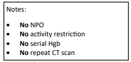
Have a look at the Regions Hospital solid organ injury protocol using the link below. It has not included serial hemoglobin levels for 18 years, which was when it was written. Take care to look at the little NO box on the left side of the page.

I’d love to hear from any of you who have also abandoned this little remnant of the past. Unfortunately, I think you are in the minority!

Reference: Serial hemoglobin monitoring in adult patients with blunt solid organ injury: less is more. J Trauma Acute Care Open 5:3000446, 2020.