All posts by The Trauma Pro

The Evolution Of Penetrating Neck Trauma Management – Part 3: Determining Risk

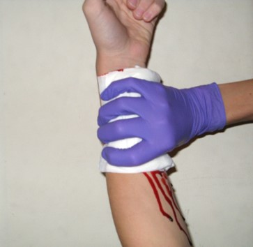
In the last post, I described the first crucial step in the contemporary management of penetrating neck trauma, control of obvious external hemorrhage. Let’s move on to the nuts and bolts of figuring out what needs to be done about the injury.

Now, it’s time to triage your patient based on clinical signs that predict the presence or absence of a significant injury. In the old days, the neck was conceptualized as three different zones that dictated the diagnostic and management algorithm.

We are now moving toward considering the neck as a single unit. The next decision point is to determine the risk for vascular or aerodigestive tract injury based on an examination for signs of injury. These signs have been divided into three groups.

Hard signs. These indicate a high risk for deeper injury and consist of the following:

  • Vascular signs
    • Refractory shock
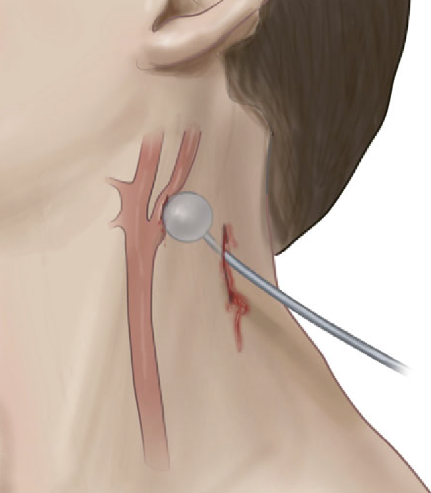
    • Pulsatile or difficult-to-control hemorrhage
    • Large or expanding hematoma
    • Audible bruit or palpable thrill (I hardly ever see anyone actually check the neck for these, so brush up your skills!)
  • Aerodigestive signs
    • Airway compromise or stridor
    • Bubbles from the wound
    • Significant subcutaneous emphysema
    • Major hematemesis
    • Massive hemoptysis
  • Neurologic signs
    • Neurologic deficits that suggest embolic strokes from a vascular injury

Soft signs. These suggest an intermediate risk for injury and are:

  • Vascular signs
    • Small or stable hematoma
    • History of bleeding or hypotension that has resolved
    • Active venous oozing
    • Pulse volume or blood pressure discrepancy (this suggests a thoracic vascular injury)
  • Aerodigestive signs
    • Hoarseness or any voice changes
    • Painful swallowing
    • Difficult swallowing
    • Mild subcutaneous emphysema
    • Minor hematemesis
    • Minor hemoptysis
  • Neurologic signs
    • Local neurologic deficit (direct injury to local nervous structures)

No signs. Obviously, this suggests a low risk of injury.

Once the level of risk has been determined, a course of action can be planned. Most patients with hard signs will require operative intervention. Plain x-rays with skin markers in place may help visualize retained foreign bodies and their relationship to bony structures. If the signs are immediately life-threatening, this step should be skipped, and operative exploration should be performed immediately. If the patient is stable and the injuries may be outside the easily accessible area of the neck (the old Zone II), a multi-detector CT angiogram (MDCTA) may help with operative planning. It may also identify patients eligible for endovascular repair.

Patients with soft signs have a lower risk of injury and should immediately undergo MDCTA. This scan has very high sensitivity and specificity in this group.

Finally, patients with no signs of deeper injury rarely need any intervention. Small series suggest that these patients could potentially be discharged from the ED. However, most trauma professionals will be uncomfortable with the thought of this. MDCTA is a low-risk test; until we know better, it’s probably best to obtain it before discharge.

Bottom line: I have described the initial assessment and management of patients with penetrating neck injury using the newer method using signs of injury in place of the old zones of injury. Nuances are still possible, such as what to do if the MDCTA is indeterminate for a vascular or aerodigestive injury. Fortunately, that is fodder for another post!

Reference: Approach to Penetrating Neck Trauma: What You Need to Know. J Trauma Acute Care Surg. 2024 Mar 25. doi: 10.1097/TA.0000000000004292. Epub ahead of print. PMID: 38523116.

The Evolution Of Penetrating Neck Trauma Management – Part 2: Initial Steps

In my previous post, I described the early days of penetrating neck injury management and introduced a paper suggesting that this concept should be revised. Today, I will summarize a paper by Siletz and Inaba that is currently in press and outlines what the contemporary way of treating these injuries should be.

Step 1. If present, rapidly control external hemorrhage and airway compromise. As always, bleeding should be controlled by direct pressure or packing. Direct pressure does not look like this:

The goal is to create a zone of pressure higher than the systolic BP perfectly in the area of bleeding. Since pressure is force per unit area, a larger area like that show above diffuses the maximum pressure and just doesn’t work. Note the ongoing bleeding shown in the picture.

Here’s what direct pressure looks like:

Or

A single finger (or maybe two) should be placed on or in the wound. If deeper bleeding is a problem, the same kind of pressure can be accomplished by packing with gauze. If gauze is used, however, pressure must usually be applied over the gauze to make sure that the underlying tissues remain pressurized.

If gauze packing is not practical because of this need for additional pressure, a urinary catheter can be inserted into the wound and inflated until the bleeding stops.

Courtesy Core EM

Airway control should ideally occur in the operating room. Given the proximity of this wound to airway structures, it is imperative that an ideal environment is present when the airway is inserted. A skilled anesthesiologist should be present, with difficult airway equipment available if needed. The surgeon should be standing by with all equipment needed to obtain a surgical airway if needed. Even though the patient may be breathing okay, the airway structures may be distorted by hematoma or injury.

You have probably noted that this is the same initial assessment we used in the old three zones approach. In the next post, I will discuss the details of a new assessment approach that considers the neck a single unit.


The Evolution Of Penetrating Neck Trauma Management – Part 1

“When the facts change, I change my mind. What do you do, sir?”

This is a famous quote from John Maynard Keynes. (Or is it? There is some debate over its authenticity, but you get the idea it tries to convey.) Our knowledge base continually changes, so we must be willing to change our minds (and practices) based on new, reliable information.

The management of penetrating neck injury is one of those facets in trauma care that has undergone slow but steady progress over the past 40 years of my career. In the old days, we quickly identified the zone of injury and proceeded to the operating room for Zone II injuries. We had to think a little harder about the other zones to be certain that we needed to be in the OR. But overall, the threshold for surgery was low.

Things have been changing. Five years ago, I published a post detailing new work by Inaba et al. at LAC+USC. This started a move toward using more straightforward criteria and advanced imaging to assist decision-making with these injuries.

In this post, I’ll summarize the original paper. In the next section, I will describe the group’s paper, which is currently in press and outlines the full framework for workup or penetrating neck injury.

The advance that makes this new method possible is based on the high degree of accuracy that CT angiography of the neck provides. It is very sensitive for identifying even minor injuries to the aerodigestive tract and vascular system.

The trauma group at LAC+USC organized a prospective, multicenter study using a multidetector CT angiography of the neck for initial screening of penetrating neck injury. This allows the evaluation of the neck as a single unit, not as three zones. It also solves the problem of trying to apply zones to injuries that cross several of them.

The new algorithm that was tested utilized an initial physical exam, first looking specifically for “hard signs” of injury.  The following were considered the hard signs:

  • Active hemorrhage
  • Expanding or pulsatile hematoma
  • Bruit or thrill over the injured area
  • Unresponsive shock
  • Hemoptysis or hematemesis
  • Air bubbling from the wound

These patients were immediately taken to the OR and explored through an appropriate incision.

Patients with no signs or symptoms were admitted and observed for at least 24 hours. All other patients were considered to have “soft signs.” They underwent multidetector CT angiography of the neck, with a scanner having at least 40 slices. Further evaluation of these patients was based on the exam and CT scan.

Here are the factoids:

  • 453 patients with penetrating neck injuries were identified during the 31-month study period
  • 9% had hard signs and were taken to the OR; 50% had soft signs and underwent CT; 41% had no signs and were observed
  • For soft sign patients, 86% of scans were negative, and all were true negatives after observation
  • 12% of soft sign patients had a positive scan, and of those, 81% were true positives
  • four patients (2%) with soft signs had too much artifact for an accurate CT, and other tests were performed; 1 of the 4 had an injury
  • Sensitivity of CTA was 100%, and specificity was 97.5% in the soft sign patients
  • The authors concluded that CTA is very reliable for identifying injuries in patients with soft signs and that patients with no signs do not require scanning, only observation

Bottom line: This was an intriguing paper that utilized both physical examination and CT angiography. The results were impressive, and they supported the argument that CTA is not required in all stable patients. With additional numbers and time, it has become clear that we can safely adopt this algorithm. My next post will flesh out the details.

Reference: Evaluation of multidetector computed tomography for
penetrating neck injury: A prospective multicenter study. J Trauma 72(3):576-584, 2012.

Video: Minimally Invasive Repair Of Rectal Injuries

Extraperitoneal rectal injury repair has evolved considerably over the past 40 years. Way back when, this injury automatically triggered exploration, diverting colostomy with washout of the distal colon, and presacral drain insertion (remember those?).

We eventually backed off on the presacral drains (pun intended), which didn’t make a lot of sense anyway. And we gave up on dissecting down deep into the pelvis to approach the injury. This only served to contaminate an otherwise pristine peritoneal cavity. Ditto for the distal rectal washout. So we have been performing a diverting colostomy as the primary method of treatment for years.

A Brief Report in the British Medical Journal Open shows us what may very well be the next stage in treating these injuries. Whereas they were previously left to heal on their own followed by colostomy closure after a few months, these authors from Sunnybrook Health Sciences Centre in Toronto are promoting a minimally invasive approach to definitive management.

They detail two cases, one an impalement by a steel rod through the rectum and bladder, and one stab to the buttock. The authors dealt with the non-rectal injuries using conventional techniques. The rectal injuries were repaired using trans-anal minimally invasive surgery (TAMIS). Both were discharged without complications.

Here is a video of the technique used in the stab victim (no audio):

video
play-sharp-fill

Bottom line: It’s about time! As long as there is not a destructive injury to the extraperitoneal rectum, this seems like a great technique to try. It may very well eliminate the need for a diverting colostomy.

But remember, this is only a case report. We don’t know about antibiotic duration, followup imaging, longer term complications, or anything really. A larger series of cases is warranted to provide these answers. This will take some time due to the low frequency of this injury. So if you try it, build your own series and publish it so we all can learn!

Reference: Minimally invasive approach to low-velocity penetrating extraperitoneal rectal trauma. Trauma Surg Acute Care Open. 2020 May 12;5(1):e000396. doi: 10.1136/tsaco-2019-000396. PMID: 32426526; PMCID: PMC7228675.

Death Knell For The IVC Filter

IVC filter insertion has been one of the available tools for preventing pulmonary embolism for decades—or so we thought. Its popularity has swung back and forth over the years and has been in the waning stage for quite some time now. This pendulum-like motion offers an opportunity to study effectiveness when coupled with some of the large datasets that are now available to us.

IVC filters have been used in two ways: prophylactically in patients at high risk for pulmonary embolism (PE) who cannot be anticoagulated for some reason and therapeutically once a patient has already suffered one. Over the years, guidelines have changed and have frequently been in conflict. Currently, the American College of Chest Physicians does not recommend IVC filters in trauma patients, and the Eastern Association for the Surgery of Trauma just released a new practice guideline for them.

A previous study from Boston University reviewed its own experience retrospectively over a 9-year period. This cohort study looked at patients with and without filters, matching them for age, sex, race, and injury severity. The authors specifically looked at mortality and used four study periods during the 9-year interval.

Here are the factoids:

  • Over 18,000 patients were admitted during the study period, resulting in 451 with an IVC filter inserted and 1343 matched controls
  • The patients were followed for an average of 4 years after hospitalization
  • Mortality was identical between patients with filters vs the matched controls

dvt-study

  • There was still no difference in mortality, even if the patients with the filter had DVT or PE present when it was inserted
  • Only 8% ever had their “removable” filter removed (!)

And now, there is a paper in press from the Eastern Association for the Surgery of Trauma with their newest practice guideline on IVC filters. They examined the literature on patients with or at risk for venous thromboembolism (VTE) and sought to determine whether IVC filters should be used prophylactically or therapeutically in these situations. They reviewed twenty-one studies, most of which were of the usual low quality.  They drew the following conclusions:

  • IVC filters should not be placed routinely for prophylaxis in patients without DVT who cannot receive chemoprophylaxis.
  • EAST conditionally recommends that IVC filters not be placed in patients with DVT who cannot receive prophylaxis. This recommendation was conditional due to the very poor quality of the few papers available to answer this question.

Bottom line: It looks like the end is near for the IVC filter. However, I can still foresee a few situations where there might be some utility. Consider the case where a patient has DVT, cannot be anticoagulated, and is showering emboli to the lungs. Otherwise, it appears that this device is on its last legs!

References:

  1. Association Between Inferior Vena Cava Filter Insertion
    in Trauma Patients and In-Hospital and Overall Mortality. JAMA Surg, online ahead of print, September 28, 2016.
  2. Role of Vena Cava Filter in the Prophylaxis and Treatment of Venous Thromboembolism in Injured Adult Patients: A  Systematic Review, Meta-Analysis, and Practice Management Guideline from the Eastern Association for the Surgery of Trauma. Journal of Trauma and Acute Care Surgery, Publish Ahead of Print DOI: 10.1097/TA.0000000000004289, 2024.