All posts by The Trauma Pro

What Does A Retained Surgical Sponge Look Like?

Surgeons and surgical residents rarely see these. And because it’s so uncommon, they frequently don’t recognize the telltale findings on radiographic studies. The TSA runs into the same problem in screening passengers for weapons and other hazards at airports. But it’s the bane of any surgeon’s existence. And it’s a major reason why OR personnel take such great pains to account for everything in the room. It is a catastrophe, and always a preventable one, when some piece of equipment goes missing and ends up left inside a patient.

A number of methods have been developed to try to eliminate this problem. They include careful counts, having someone record anytime anything is placed inside, x-rays, and most recently, RFID tags.

After counting, x-ray is the most common way to try to find missing objects. One would think that these foreign bodies would be easy to see. Metallic instruments are rather easy to spot. But many trauma professionals, even those who work in the OR, have never seen what a positive image of a sponge actually looks like. So here they are. You should never miss one on an xray now.

Surgeons typically use two types of sponges in the OR: Ray-Tec sponges and standard lap pads. Ray-Tecs look like a 4×8 piece of gauze with a mysterious blue string woven throughout it. The string is the only part that shows up on x-ray, and it is very thin and somewhat hard to see. Here are some Ray-Tec sponges outside the body:

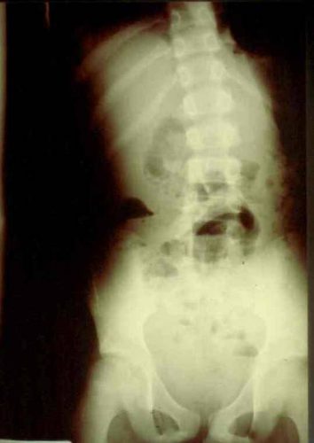
And here’s one that was left inside. Note the little squiggle in the left lower quadrant and how easy it is to overlook.

On the other hand, a laparotomy pad is a 4×4 folded cloth pad that unfolds into a larger pad. It has a blue radiopaque tag sewn in the corner, extending along one edge of the pad. Here’s what they look like outside the body:

And here’s one inside a patient. Note the irregular object in the right upper quadrant. Many times the tag is scrunched up and doesn’t look like one.

Bottom line: It’s important for anyone who works in the OR on any body part to be familiar with the appearance of these tags on x-rays. Since it’s generally impossible to get accurate counts before or after a trauma procedure, always image the involved body cavity looking for these telltale signs before closing the patient.

Note: These images  were taken from the internet. Patients were not treated at Regions Hospital.

Chest Tube Size Doesn’t Matter?

It’s great when you read a study that supports your own biases. But it’s not pleasant at all when you find one that refutes what you’ve been teaching for years. Well, I found one of those and I wanted to share it with you.

I’ve always said that there are only two sizes of chest tube for trauma, big (36Fr) and bigger (40Fr). Although there was never any good literature, it seemed intuitive that a large tube would help ensure drainage of bigger clots if hemothorax was present.

A multicenter observational study was carried out that looked at 353 chest tube insertions. This work monitored retained hemothorax or pneumothorax, the need for tube reinsertion or invasive procedure due to incomplete drainage, and pain during insertion.

Here are the factoids:

  • There was roughly a 50:50 large (36-40Fr) vs small (28-32Fr) mix of chest tubes
  • Tubes inserted for hemothorax were also a 50:50 mix of large vs small
  • The initial amount of blood out was small and about the same for both groups
  • There was no significant difference in pneumonia, retained hemothorax, or empyema
  • The need for an invasive procedure (VATS or thoracotomy) was about 11% in both groups
  • Interestingly, there was no difference in visual analog pain score between the groups either.

Bottom line: Basically, large tube and small tube were the same. So maybe chest tube size selection doesn’t matter as much as we (I?) think. It seems to make sense to select a tube size based on your patient’s chest wall, not dogma. Although subjective pain seems to be the same as well, pain and sedation management are key because this is not a fun procedure for the patient, regardless of tube size. I’m not fully convinced yet, and would like to see an additional confirmation study if possible.

Reference: Does size matter? A prospective analysis of 28–32 versus 36–40 French chest tube size in trauma. J Trauma 72(2):422-427, 2012.

What The Heck? Pigtail Catheter Chest Tube – The Answer

I previously described a trauma patient who had a pigtail type chest tube inserted with some odd CT findings after insertion:

So what is wrong in this picture? Well, the catheter has been inserted into the spleen! This can occur if it is inserted too low, or if there are adhesions between lung and chest wall or diaphragm.

How can it be avoided? Make sure that the insertion point is no lower than the 5th intercostal space. This is the level of the nipple in a male. And depending on what type of kit you use, be careful! Some are based on Seldinger technique, which would seem to be a bit safer. Others use a small trochar, which can be inserted a little too deeply at times. Note that this complication can occur with any kit, and can also occur when using a standard tube and open insertion technique.

Does a pigtail tube even work for hemothorax? There’s some debate about this. Traumatic hemothorax is not defibrinated like a medical one. Thus, there are frequently clots present which may not fully evacuate through a standard chest tube, let alone a tiny one. Thus, I don’t recommend a pigtail for acute traumatic hemothorax.

How should I manage this issue? Obviously, this tube needs to come out. And assuming that the initial indication for the tube is still present, a better one needs to be inserted. Dont’ pull it out yet! First, look at the vital signs. If there is significant bleeding and/or vitals are not normal, an immediate trip to the operating room is in order. In this case, the patient will likely lose their spleen.

If vital signs are stable, book both an interventional radiology suite and an OR. Or better yet, use a hybrid room. Have the radiologist obtain a baseline angiogram, and position a catheter in the main splenic artery. Incrementally remove the pigtail, hand injecting a small amount of contrast each time. If extravasation is noted at any time, the radiologist can then attempt to embolize. If selective embolization isn’t successful, then the main splenic artery should be embolized. If embolization doesn’t work, or vital signs deteriorate at any time, the surgeon should immediately proceed to laparotomy. Attempts at splenic salvage will probably not be successful.

Finally, insert a new, conventional chest tube using finger guidance. Don’t make the same mistake twice! And by the way, this works for pigtails in the liver, too. They are less likely to bleed significantly when withdrawn, and obviously the radiologist can only used selective embolization if they do.

What The Heck? Pigtail Catheter Chest Tube

Here’s a case to make you think!

A patient arrives after being t-boned in his driver side door. He complains of left sided chest and abdominal pain. Chest x-ray shows a modest left hemopneumothorax. The decision is made to insert a pigtail type chest tube, and this is carried out in your trauma bay. It is uneventful, and a small amount of blood but no air is returned. The pelvis x-ray is unremarkable

The patient is then taken to CT, where an abdomen/pelvis scan with contrast is performed. This interesting slice is noted. What the heck?!

Here are my questions:

  • What is wrong in this picture?
  • How could it have been avoided?
  • Does a pigtail chest tube work for hemothorax?
  • How should this issue be managed, and where?

I’ll address these questions in my next post, and more!

Image source: internet

An Audit Tool For Your Massive Transfusion Protocol

Every trauma center is required to have a massive transfusion protocol (MTP). This protocol lays out in precise detail how large quantities of blood products get to and into your patient when needed. It’s important to have all of these processes worked out in advance so that the products are safely and rapidly available.

But what happens after the MTP winds down is equally important. Without a detailed analysis of the entire process, it’s impossible to know if all of its components worked as planned. While a few centers activate the MTP frequently enough to be smooth and well-practiced, many do not. For those, it’s even more critical to pick each activation apart, looking for ways to improve.

Here are some of the important things to review:

  • Demographics
  • Components used for ratio analysis
  • Lab values (INR, TEG, Hgb)
  • Logistics
  • Waste

Bottom line: I’ve included links to two audit tools below. The Broxton tool is more rudimentary, but is a good start. The Australian tool is excellent, in my opinion. It covers all the bases, and allows the center to get meaningful information and/or research material from the data.

Do you have a great MTP audit tool? Please send me a copy so I can share.

Related posts: