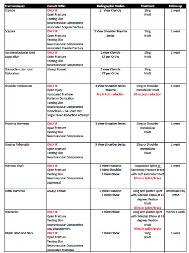
And here’s the last in my short “When To Call” series. This one’s a little different, and quite a bit longer. That’s due to the complexity and sheer number of potential orthopedic problems.
When consulting a specialty service, always keep the patient paramount in your decision making. Then think about how soon and under what context they really need to see the patient. Can it wait until morning? Do they even really need to be seen in the ED, or can this be an outpatient visit?
Tomorrow: Expectations on how your consultants should go about their business when seeing your patients.
Here’s another in my series of “When To Call” pieces. We sometimes overuse our consultants and call then at inappropriate times. So what if we diagnose an injury in their area of expertise at 2 am? Does it need attention or an operation before morning? If not, why call at that ungodly hour?
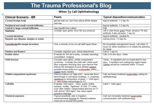
Let’s use our consultants wisely! I’ve listed most of the common eye diagnoses that trauma professionals will encounter. There is also an indication of what you need to do, and exactly when to call your consultant.
Unfortunately, this one won’t fit on a 3×5 index card that you can keep in your pocket. I’ve included a printable pdf file, as well as the original Microsoft Word file in case you want to make a few modifications to suit your own hospital.
In my next post, I’ll provide a comprehensive reference for when to call your orthopedic surgeon.
Here’s the first in a series of “When To Call” pieces. We sometimes overuse our consultants and call then at inappropriate times. So what if we diagnose an injury in their area of expertise at 2 am? Does it need attention or an operation before morning? If not, why call at that ungodly hour?
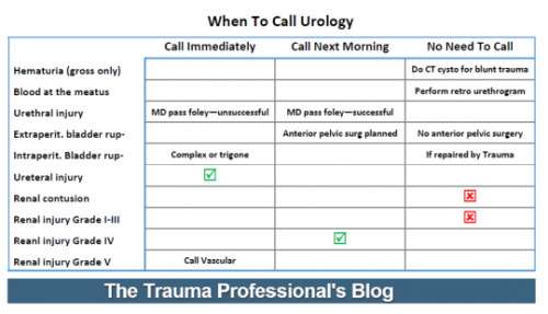
Let’s use our consultants wisely! I’ve listed most of the common urologic diagnoses that trauma professionals will encounter. There is also an indication of what you need to do, and exactly when to call your consultant.
Here’s a reference sheet formatted at a 3×5 index card that you can keep in your pocket. I’ve included a printable pdf file, as well as the original Microsoft Publisher file in case you want to make a few modifications to suit your own hospital.
Tomorrow, a reference card for your eye consultant.
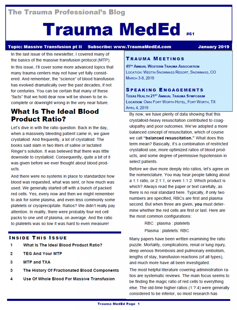
Welcome to the current newsletter. This is part 2 of my discussion of the massive transfusion protocol (MTP). Here are the topics I cover:
What Is The Ideal Blood Product Ratio?
TEG And Your MTP
MTP and TXA
The History Of Fractionated Blood Components
Use Of Whole Blood For Massive Transfusion
The next issue covers fat embolism syndrome and will be released to subscribers at the end of the month. Non-subscribers will have to wait another week for the public release.
The scenario involved an elderly woman who fell from standing at her care facility 12 hours earlier. They want to send her to your trauma center for evaluation because she seems a bit different from her baseline. You have well defined practice guidelines for patients with head injuries that dictate what type of monitoring and diagnostics they receive.
What do you need to know to determine what you should do? Thanks for all of you who sent in suggestions.
Here are my thoughts:
Which scans should she get? Usually, you would obtain an initial head CT and, due to her age, a cervical CT regardless of her physical exam due to the high miss rate in these patients. But now the fun begins. Your subarachdoid / intraparenchymal hemorrhage (IPH) practice guideline would have you admit for neurologic monitoring for 12 hours, obtain a TBI screen, then discharge without a followup scan if the screen was passed. But in this case, the clock started 12 hours ago and the guideline would be finished with the exception of the TBI screen. So an initial scan and a TBI screen in the ED are all that are needed. The observation period is already over and the patient could potentially be discharged from ED if a SAH or IPH were found.
Your subdural guideline mandates all of the above plus a repeat scan at 12 hours. But once again, the clock has already started. Do you just get an initial scan, which also serves as the 12 hour scan? Or do you get yet another one? If the neuro exam is normal, I vote for the former, and your evaluation is complete after the TBI screen. If the neuro exam is not quite normal, then admission for continuing exams and a repeat scan are in order.
Does the patient need to be admitted, and for how long? Hopefully, you’ve figure this out in the previous bullet. The clock started running when she fell down, so in cases where the physical exam is normal, only the first CT is needed and ongoing monitoring is not. Thus, she could return to her care facility from the ED after the scan.
What other important information do you need to know? Of paramount importance is her DNR status and her/her family’s willingness to have brain surgery if a significant lesion is identified. It is extremely important to know the latter item. If there is never any patient or family intent to proceed to surgery, is there any point to obtaining scans at all? In my opinion, no. The whole reason to obtain the scan and monitor is to potentially “do something.” But if the patient and/or family will not let us “do something,” there is no reason to do any of this. It is crucial that the patient and family understand the typical outcomes from surgery given her age and degree of frailty. This is most important in patients who are impaired with dementia or a high-grade lesion if found from which there is minimal chance of recovery. In most such cases, even if surgery is “successful,” the patient will never recover enough to return to their prior level of care. This should be weighed heavily by the family and care providers.
Should a patient with DNR or “no surgery” orders even be sent to the ED? Theoretically, no. There is no need from the standpoint of their future care. They are not really eligible to have any studies or monitoring done. However, the facility may try to insist for their own liability issues, but this is not really a valid clinical reason.
I hope you enjoyed this little philosophical discussion. Feel free to agree/disagree through your comments or tweets!
Home of the Trauma Professional's Blog
Do you want to get a daily email every time there’s a new post? See what I’m up to.