All posts by The Trauma Pro

Best of AAST #7: What’s New With Reboa

Despite all you read about it these days, REBOA is still very new. The first papers describing use in humans are barely 5 years old! A few select centers have been early adopters and are publishing a regular flow of research on their experience.

But we need more numbers! Many trauma centers have considered, or actually adopted the use of REBOA already. However, we are still working out a lot of the nuts and bolts of this very invasive procedure. The group at University of Arizona – Tucson reviewed the national experience over a two year period by massaging the data in the Trauma Quality Improvement Program (TQIP) database. All Level I-III trauma centers in the US are required to report their experience to this large, detailed collection of trauma data.

They performed a retrospective review of REBOA vs non-REBOA patients matched for demographics, prehospital and emergency department vital signs, mechanism of injury, degree of pelvic disruption in pelvic fracture patients, solid organ injuries, and lower extremity fractures and vascular injuries. The studied outcomes were complications and mortality.

Here are the factoids:

  • Nearly 600,000 records were scanned for the two year period, and only 140 REBOA patients were identified (!)
  • These 140 REBOA patients were matched with 280 similar non-REBOA patients
  • Average age was 44 and average ISS was 29, 74% were males and 92% were blunt trauma
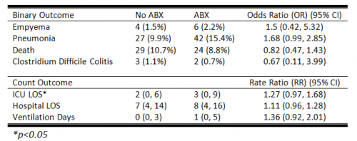
  • Overall complication rate was 7.4% and mortality was 25%
  • There was no difference in 4-hour or 24-hour numbers of blood, plasma, or platelets transfused
  • ICU and hospital length of stay were identical
  • 24-hour mortality in the REBOA group was significantly higher (36% vs 19%)
  • REBOA patients were significantly more likely to require amputation (5% vs 1%)

Bottom line: These are not great numbers for REBOA! What gives? There are a number of possibilities:

  • It’s a database study, so some key information might be missing
  • The numbers remain small, only 140 patients out of half a million records in two years!
  • There is no way to know how the patients were selected for REBOA
  • The experience and skill level at the hospital performing the procedure is not known
  • The interplay of other injuries and comorbidities is unclear
  • And many more…

BUT, the numbers are concerning. The early adopter centers have better outcomes, and this has prompted many centers with fewer eligible patients to jump on the bandwagon. We all need to remember that this is a brand new procedure and we are still learning the nuances. It is extremely important that every center performing REBOA contribute their results to a national registry. We still need to figure out which patients will benefit from it, how it should be used, and how we can minimize complications and maximize survival in our patients.

Reference: Nationwide analysis of resuscitative endovascular balloon occlusion of the aorta (REBOA) in civilian trauma. Session I Paper 5, AAST 2018.

Best of AAST #6: Antibiotics For Chest Tubes??

For as long as I can remember (nearly 50 years worth of literature) there has been some debate about giving antibiotics after chest tube insertion to decrease the infection rate. The pendulum moved back and forth for decades, never getting very far into the “give antibiotics” side. It’s been quite a while since I remember any new papers on this, and I thought the debate had been resolved in favor of never using them.

But then I see an abstract from the AAST multi-institutional trials group studying presumptive antibiotics after chest tube insertion! They conducted a prospective, observational study at 22 Level I trauma centers, enrolling nearly 2,000 patients. They matched patients in antibiotic and no antibiotic groups, arriving at (only) 272 patients in each group.

Here are the results:

Bottom line: First, it’s a little disappointing that the numbers were so low with a trial that includes 22 trauma centers. Did they have a hard time finding centers that would give antibiotics? Or was it just hard to match patients for the variables they were looking at? Regardless, there were no significant differences in infectious complications, and a non-clinically significant difference in ICU stay with antibiotics.

Why won’t this die? If there are so few papers that show an actual benefit from giving antibiotics after chest tube insertion with 50 years of data, then it’s very unlikely that it will ever be shown to be necessary!

Reference: Presumptive antibiotics for tube thoracostomy for traumatic pneumothorax. Session XXII Paper 49, AAST 2018.

Best of AAST #5: Pneumothorax – How Big Is Too Big?

Deciding when to place a chest tube can be challenging. Sometimes, it’s obvious: there is a large hemo- or pneumothorax staring you in the face on the chest x-ray. But sometimes, it’s there but “not that big.” The real question is, how big is too big.

That’s a question that’s been very difficult to quantify. The authors of this abstract, from the Medical College of Wisconsin, conducted a six-year retrospective review of every patient with an isolated pneumothorax at their Level I trauma center. Based on their previous research, a 35mm threshold was used to stratify patients into two groups. This measurement was obtained from axial images of a CT scan. Statistical analysis was performed to identify the predictive value in determining whether the patient could be managed without a chest tube.

Here are the factoids:

  • A total of 1767 patients had a pneumothorax during the 6-year period, and about half met inclusion criteria for the study
  • Of the 385 with pneumothorax alone, 92% were managed without a chest tube
  • Of those 353, 95% had a maximum chest wall to lung distance (335)
  • The 35mm measurement was statistically shown to be an independent predictor of successful management without a tube for both blunt and penetrating trauma

Bottom line: Not so fast! Although this looks like a slam dunk abstract, it’s really not. First, many (or most?) pneumothoraces are initially diagnosed using a plain old chest x-ray. A 35mm measurement is meaningless here because there can be significant changes in position of the pneumothorax on the image. Sometimes, the air is located anteriorly with little or no lateral component.  Does this mean we should CT every patient with a known or suspected pneumothorax? I think not.

And the second issue is the subjectivity surrounding the definition of a failure. What criteria were used when the tube was actually placed in this series. If every patient had to become symptomatic first, then I might agree. But I suspect the tubes were placed when followup imaging showed that the air was just “too big.” You can’t statistic away this kind of potential bias from subjectivity.

So what’s the answer? Unfortunately, there still isn’t one. The need for a chest tube must still be based on subjective size on a chest x-ray, physiologic status, and the patient’s ability to tolerate a given amount of lost lung function. It continues to boil down to the  assessments of each trauma professional as to “how big is too big.”

Reference: Observing pneumothoraces: the 35mm rule is safe for both blunt and penetrating chest trauma. Session XVA Paper 28, AAST 2018.

Best of AAST #4: Better Triage For Scene Helicopter Transport

There has been tremendous debate around the value and use of prehospital helicopter emergency medical services (HEMS). It’s fast, but also expensive, and there is always a small amount of added risk to patients during transport. Over the years, there has been a significant increase in the number of helicopter services, and in some cases it seems like several services are dashing to accident scenes in the hope that they can pick up the patient.

Overuse of HEMS has also been recognized, with some patients transported who could have just as easily and safely been moved by ground ambulance. This is a particularly vexing problem with pediatric patients.

The holy grail of trauma HEMS has been to find some easy to identify scene variables that reliably predict which patients should be transported by air. A group in North Carolina tapped the state trauma registry to attempt to develop such a system. They analyzed data in the registry over a three year period, mathematically analyzing for easily identified predictors of ED death or need for operating room, interventional radiology, or ICU admission.

Here are the factoids:

  • The percentage of flights from the scene increased from 7% to 9% compared to data from fifteen years prior to this study
  • Vital signs (SBP, pulse, GCS motor) had the best correlation with mortality, and these were used to develop a regression model for triage
  • Patients with normal SBP, pulse, and GCS motor of 6 were found to safely transported by ground EMS, with similarly low mortality for ground or air
  • During the study period, triaging patients that met these criteria would have saved the state system about $19 million

Bottom line: Every state should take a look at their guidelines for helicopter vs ground transport for scene runs. This is an expensive tool, and should be treated with respect. Just because a helicopter is available does not mean it should be used. The commander on the scene must make the proper decision based on variables like these, but also apply their knowledge of traffic patterns, time and distance from the most appropriate receiving trauma center.

Reference: Trauma system resource preservation: a simple scene triage tool can reduce helicopter emergency medical services (HEMS) over-utilization in a state trauma system. Session IV Paper 13, AAST 2018.

Best of AAST #3: Level I vs Level II Trauma Centers

There is an ongoing debate over the differences between Level I vs Level II trauma centers in the US. On paper, the major differences include resident rotations in trauma, research, and the available of certain specialty surgeons and services. There have been several papers that look at survival differences between the two levels.

One podium paper at AAST 2018 re-examines this debate. It is a medium-sized pooled series that looks at a particular type of injury, pelvic ring fractures. These injuries can be complex, and many times require specialized orthopedic expertise. ACS Level I centers are required to have at least one Orthopedic Trauma Association fellowship-trained surgeon among their orthopedists. This is not required for Level II centers, but many do have them.

The group at the University of Michigan examined patients with partially stable or unstable pelvic ring injuries in a trauma collaborative database including 29 Level I and Level II centers over a 7-year period. They used propensity matching to compare 610 patients admitted to Level I and 610 patients admitted to Level II centers with these injuries:

Here are the factoids:

  • Mortality was significantly increased at Level II centers ( 12%) vs Level I centers (8%)
  • Angiography was used significantly less at Level II centers (6% vs 11%)
  • Complex repairs were used significantly less frequently at Level II centers (32% vs 42%)
  • Patients were significantly less likely to be admitted to an ICU at Level II centers, and were more often admitted to stepdown units (45% vs 52%)
  • Failure to rescue rate was lower (better) in ICU patients

Bottom line: Obviously, there are some limitations to using this pooled data, but it does provide larger numbers than many similar papers have. It cannot distinguish Level II centers that have OTA-trained orthopedic surgeons from those that do not. But the results are rather striking. It’s not clear exactly which of the institutional differences might be responsible for the improved mortality, and they all probably contribute to some degree. But the abstract appears to show that Level II centers are not just non-academic Level Is. This work suggests that certain injury patterns really should be transferred to a center with the specialized resources to treat it well.