Best Of EAST #7: King vs. I-Gel Supraglottic Airways

Airway assessment and protection are of paramount importance during trauma care. Airway management is even more challenging in the prehospital environment, where lighting and patient positioning may be suboptimal, and injuries or policies may prohibit orotracheal intubation. A variety of devices have been developed to make airway control simpler and faster.

Both the King and i-gel airways were introduced around 2005. The former functions as an extraglottic airway, and the latter as a supraglottic one. Prehospital providers typically use these devices for airway control across the US. They are both designed for blind insertion without the need for other equipment, such as a laryngoscope.

The group at West Virginia University compared these two devices based on the incidence of hypoxia, cardiac arrest, successful insertion, and survival to hospital discharge. They performed a large database search and attempted to control for patient age, weight, pre-airway hypoxia, and the use of suction.

Here are the factoids:

  • A total of 1,557 patients were studied; one-third received a King airway, and two-thirds had i-gel insertion
  • Half of all patients experienced hypoxia, and a quarter experienced severe hypoxia (saturation < 80%) after insertion of any tube
  • But i-gel placement was not associated with hypoxia, severe hypoxia, cardiac arrest, or decreased survival to discharge, and had better success on first-pass placement

Bottom line: What? This is a first. I honestly can’t figure this abstract out. The two bullets in red above cancel each other out. If half of all patients had hypoxic episodes, and only one-third had King airways, that means that at least 16% of the i-gel patients experienced hypoxia. I can’t reconcile that with the last bullet, where i-gel was not associated with any adverse events.

Several other papers have compared the use of these two devices over the last two decades. Most suggest that the i-gel is simpler, with fewer misplacements and other complications, and tends to be preferred by prehospital personnel.

Unfortunately, I have to disregard this entire abstract due to the conflicting data listed. Perhaps the presenter will clarify or provide some corrections to the data. Otherwise, I have not learned anything from it, and it doesn’t appear to add anything new to the trauma literature.

Reference: A retrospective comparison of the King laryngeal tube and i-gel supraglottic airway devices for injured patients. EAST 2024 Podium paper #27.

Best Of EAST 2024 #6: Pan-Scans In the Elderly

Injured older adults typically sustain those injuries from blunt mechanisms. Radiographic evaluation, particularly CT scans, does not have good supporting literature to dictate which exams should be used in particular patients. There is a long-standing debate on the merits of pan-scan vs. selective scans when using CT.

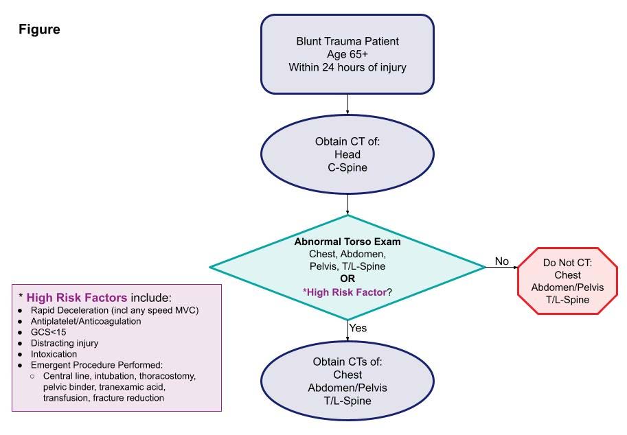
EAST sponsored a multicenter study to look for specific history and physical exam findings that could help direct CT evaluation. Eighteen Level I and II trauma centers participated in a prospective study of patients aged 65 and above. The authors used machine learning to determine clusters of findings that could be used to create decision rules for using a pan-scan vs a tiered scan (head + cervical spine, then possibly torso). The focus was on injuries to the head/cervical spine and the torso. Patients who presented late after injury (24 hours) or died were excluded, leaving 2,587 for study.

Here are the factoids:

  • The learning system could not develop a rule for pan-scan usage
  • A high-quality model was created to guide the use of selective scanning, which was 94% sensitive with an 86% negative predictive value

  • The authors estimated that 12% of patients would be spared a torso scan using the decision tool

The authors concluded that their decision tool has promising sensitivity and negative predictive value and needs further prospective evaluation to validate it.

Bottom line: As our use of machine learning and AI advances, work like this will continue to accelerate. One of the benefits of using AI as a tool is its ability to sift through enormous amounts of raw data to detect faint but significant signals. 

The downside is that much of what we record is garbage,  making detecting that signal much more difficult. However, the process is inexpensive and can potentially advance health care in a variety of ways. 

The presenter and authors should describe the specific machine learning technique used and outline its particular strengths and weaknesses. Expect to see more and more abstracts like this coming down the pike. And when someone starts applying large language models like Chat-GPT to this type of medical data, look out!

Reference: Scanning the aged to minimize missed injury: an EAST multicenter study. EAST 2024, Podium paper #24.

Best Of EAST #5: How Good Is Lung Ultrasound For Pneumothorax?

Focused abdominal sonography for trauma (FAST) has been a mainstay of rapid diagnosis for many years. The extended FAST exam (eFAST) adds an examination of the thoracic cavities to the basic exam. The sensitivity and specificity of FAST have mostly been determined. However, there is much less literature outlining the accuracy of the eFAST.

The group at Vanderbilt performed a prospective, observational study on the ability of the eFAST exam to detect pneumothorax specifically.  Imaging was performed by a licensed sonographer and read by the attending surgeon and a radiologist as it was completed.

Here are the factoids:

  • Only patients receiving chest X-rays, chest ultrasound, and chest CT for confirmation were included in the study, which totaled 1,499
  • The statistical analysis was as follows:
  • Only 25% of patients had a chest tube placed
  • Patients with false negative exams were more likely to have rib fractures and lower oxygen saturation and blood pressure

The authors concluded that chest ultrasound had a low sensitivity and high false negative rate and that many of the negatives eventually required chest tube insertion. They advise that the chest ultrasound should be used alone with caution.

Bottom line: This is an interesting and relatively large study. However, it is at odds with a paper published in 2021 from George Washington University. They published a series of 3,410 patients and found 71% sensitivity, 99% specificity, 87% PPV, and a 2.2% false negative rate. 

These are very disparate numbers. However, the GW study was retrospective and included both pneumothorax and fluid in the abdomen or chest, whereas this abstract looked strictly at pneumothorax and was prospective.

Both studies are interesting on their own, and the presenter of this abstract will need to explain why the results are so different.  The answer to the question of how much we believe the eFAST result remains up in the air!

References:

  1. Lung ultrasound underdiagnoses clinically significant pneumothorax. EAST 2024, Podium paper 21.
  2. eFAST exam errors at a level 1 trauma center: A retrospective cohort study. Am J Emerg Med. 2021 Nov;49:393-398.

Best Of EAST 2024 #4: Chest Tube Irrigation Prevents Retained Hemothorax

One of the potential complications of chest trauma causing hemothorax is the retained hemothorax. In most patients, retained blood slowly lyses and is reabsorbed. But a few do not, and scarring can occur that entraps the lung and interferes with pulmonary function. This can ultimately require a VATS or thoracotomy to resolve.

Several protocols have been developed to try to prevent a retained hemothorax. They include the use of lytics or an early VATS procedure. The group at the Medical College of Wisconsin performed a trial of thoracic cavity irrigation and compared the outcomes with patients who did not undergo irrigation. This was a single-center retrospective study performed over five years.

This appears to have been a common practice at this institution. Patients undergoing chest tube placement for hemothorax received irrigation of the chest cavity immediately after placement. The study excluded patients with chest tubes placed in other hospitals, tubes placed late (after 24 hours), or patients who had a chest procedure within 6 hours.

Here are the factoids:

  • A total of 370 patients were enrolled, and 225 (61%) received irrigation
  • Demographics of the groups were the same, with the exception that the irrigation group contained more patients with penetrating injury and fewer patients with flail chest
  • Use of irrigation was associated with significantly less incidence of retained hemothorax (10% vs 21%) or need for VATS (6% vs. 19%)
  • Chest tube duration (4 vs 6 days) and hospital length of stay 8 vs 10 days) were also significantly shorter

The authors concluded that irrigation prevents retained hemothorax and decreases the need for surgical intervention.

Bottom line: Well, this was a new one for me. The only prior study I could find was published in 2022 by a group at the University of Nevada at Las Vegas. They irrigated 82 of 198 patients undergoing chest tube placement. They noted a decrease in hospital, ICU, and ventilator days.

This study looks at something far more practical: interruption of the development of a complication. Although still a relatively small and single-institution study, it was well done and could easily detect statistical significance. 

The presenter should be prepared to discuss what impact the mechanism of injury (penetrating, flail chest) may have had on their results and the exact technique they used. How much fluid, what type, and how it was drained are all important questions to discuss.

This is a fascinating abstract indeed. If the presentation answers the questions, centers should consider updating their chest tube management algorithms.

References:

  1. Thoracic cavity irrigation prevents retained hemothorax and decreases surgical intervention in trauma patients. EAST 2024, Podium paper #17.
  2. The Volume of Thoracic Irrigation Is Associated With Length of Stay in Patients With Traumatic Hemothorax. J Surg Res. 2022 Nov;279:62-71.

Best of EAST 2024 #3: VTE Prophylaxis For Pediatric Trauma

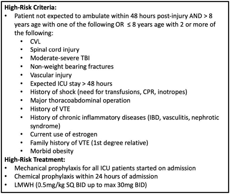
Venous thromboembolism (VTE) after trauma in adults has generated a considerable body of literature for guidance. However, there is much less information available regarding pediatric trauma. High-risk criteria for pediatric VTE after trauma have recently been released.

These criteria have not yet been evaluated prospectively or coupled with the administration of chemoprophylaxis. The Medical College of Wisconsin trauma group organized a prospective, multi-institutional study involving eight pediatric trauma centers. They studied VTE events within 30 days and bleeding complications. The children were stratified into three groups: no prophylaxis, early prophylaxis (within 24 hours), and late prophylaxis.

Here are the factoids:

  • A total of 460 patients were enrolled during a three-year period
  • The number of VTE events was very low at 25 (5.4%)
  • Patients who developed VTE had a median of 4 of the high-risk criteria, most commonly ICU stay>48 hours and TBI.
  • Half of patients received prophylaxis
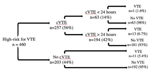
  • VTE occurred in 1.6% receiving an early dose and 6.7% with late dosing
  • There were no bleeding complications

The authors concluded that prophylaxis in children at high risk for VTE was safe, but they could not demonstrate any risk reduction for those who had received chemoprophylaxis compared to those who had not.

Bottom line: This is another study that was cursed by low numbers. See the breakdown chart below:

There is a trend toward higher VTE in children receiving prophylaxis late or never. However, the number of subjects is far too low to detect significance. The good news is that there were no bleeding events in this modest sample of 257 patients. 

So what next? The authors state that “further subgroup analysis is ongoing to refine the high-risk criteria.” Good luck with that because subgrouping will deplete the numbers even further.

There are several things the authors could do to improve this work:

  • Get more subjects! Increase the number of centers participating, and consider sending it through the EAST Multicenter Trial process.
  • Streamline the list of high-risk criteria. There are quite a few of them. Try to focus on the most obvious ones and make sure each one has clear definitions. And set a threshold of how many must be present to trigger chemoprophylaxis.
  • Define the pediatric patient precisely. As children approach puberty, they behave more like adults as it pertains to VTE. State an explicit age cutoff.

This presentation should be a springboard to soliciting help from other pediatric trauma centers so this group can return to this meeting with compelling information.

Reference: The No Clot VTE study in high-risk pediatric trauma patients. EAST 2024 Podium paper #6.