Chest Tube Repositioning – Final Answer

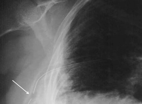
So you’re faced with a chest tube that “someone else” inserted, and the followup chest xray shows that the last drain hole is outside the chest. What to do?

Well, as I mentioned, there is very little written on this topic, just dogma. So here are some practical tips on avoiding or fixing this problem:

  • Don’t let it happen to you! When inserting the tube, make sure that it’s done right! I don’t recommend making large skin incisions just to inspect your work. Most tubes can be inserted through a 2cm incision, but you can’t see into the depths of the wound. There are two tricks:
    • In adults with a reasonable BMI, the last hole is in when the tube markings show 12cm (bigger people need bigger numbers, though)
    • After insertion, get into the habit of running a finger down the radiopaque stripe on the tube all the way to the chest wall. If you don’t feel a hole (which is punched through the stripe), this will confirm that the it is inside, and that the tube actually goes into the chest. You may laugh, but I’ve seen them placed under the scapula. This even looks normal on chest xray!
  • Patients with a high BMI may not need anything done. The soft tissue will probably keep the hole occluded. If there is no air leak, just watch it.
  • If the tube was just put in and the wound has just been prepped and dressed, and the hole is barely outside the rib line, you might consider repositioning it a centimeter or two. Infection is a real concern, so if in doubt, go to the next step.
  • Replace the tube, using a new site. Yes, it’s a nuisance and requires more anesthetic and possibly sedation, but it’s better than treating an empyema in a few days.

Chest Tube Repositioning – Part 2

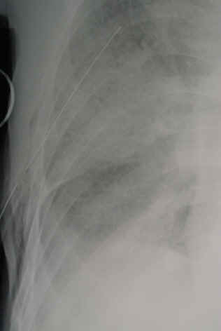
Yesterday I presented the problem of the malpositioned chest tube, specifically one that is not completely in the pleural space. This one is way out:

So what do the doctor books say? Well, the first thing you will discover if you try to look it up is that THERE IS NO LITERATURE ON THIS COMMON PROBLEM! There are a few papers on tubes placed in the fissure and tubes inserted into the lung parenchyma. But there are only a few mentions of tubes with holes still outside the chest.

I’ve gotten a number of comments, including “you can push them in a little”, “take it out and put in another”, and “never push them in.” Since we don’t have any science to guide us, we have to use common sense. But remember, I’ve shown you plenty of examples where something seems reasonable, but turns out to be ineffective or downright harmful.

There are three principles that guide me when I face this problem:

  • Prevention is preferable to intervention
  • Do no (or as little as possible) further harm
  • Be creative

Tomorrow, I’ll finish this series and provide some tips and guidelines to help manage this problem using the principles outlined above.

What To Do When The Chest Tube Is Not In The Right Place

It happens from time to time. Your patient has a hemothorax or pneumothorax and you insert a chest tube. Well done! But then the xray comes back:

The last hole in the drain is outside the chest! What to do???

Here are the questions that need to be answered:

  • Pull it out, leave it, or push it in?
  • Does length of time the tube has been in make a difference?
  • Does BMI matter?

Leave comments below regarding what you do. Hints and final answers in my next post!

Can We Use Type A Plasma For Emergency Transfusion?

Trauma patients tend to try to bleed to death. And trauma professionals try to stop that bleeding. They also frequently have to replace the blood products that were lost, which includes red blood cells, plasma, platelets, and more.

From a red blood cell standpoint, we have a long history of using group O- packed red cells as the so-called universal donor product. The problem is that only about 5% of the world population has this blood type, so it can be scarce.

To address this, many centers have moved toward using O+ blood for select patients. This blood type is much more prevalent (about 50% worldwide). The only difference is the positive Rh factor which has little impact on males, or females who are not in their child-bearing years. If an allergic reaction occurs, it is typically mild.

But what about plasma? This is interesting stuff. When selecting red cells, we want them to have no ABO group antigens on them so they don’t provoke a reaction. But plasma is just the opposite. We don’t want any ABO group antibodies in it. And the only plasma without antibodies comes from people who have all of them (A and B) on their red cells. This means people with type AB+ blood. Unfortunately, this is the other rare blood type, so there’s not a lot to go around. Worldwide, about 5% of people are AB+ and less than 1% are AB-.

So why couldn’t we do something like we did with packed red cells and substitute a more common blood type that evokes little immune response? The American Association of Blood Banks (AABB) has authorized both AB and A plasma for use in emergency situations. Unfortunately, the safety profile for using group A has not been very well studied, particularly in trauma patients needing massive transfusion.

The authors of the PROPPR study re-analyzed the data from it to try to answer this question. As you may recall, PROPPR was published in 2015 and compared safety and effectiveness of transfusion ratios at 1:1:1 to 1:1:2 (plasma : platelets : red cells).

The study group selected patients from the dataset who received at least one unit of emergency release plasma (ERP), defined as product given before the patient’s ABO type had been determined. Nicely enough, 12 sites transfused group AB ERP and 9 sites gave group A. One site gave both A and AB.

The authors looked at in-hospital mortality at 30 days, and a host of complications. Here are the factoids:

  • A total of 584 of the 680 patients in the PROPPR study received emergency release plasma
  • The median number of units given was 4, and there was no difference between A and AB groups
  • There were statistically significant baseline differences between the groups, including blood type, SBP, percent in shock (SBP<90), blunt mechanism, positive FAST that were probably not very clinically significant
  • The number of transfusions of all products were significantly  higher in the A plasma group
  • Complications were significantly higher in the A plasma group, specifically from SIRS, pulmonary problems, and venous thromboembolism (VTE)
  • There were no acute hemolytic transfusion reactions and three febrile reactions

The authors concluded that, statistically, the use of group A plasma was not inferior to the use of group AB. The authors stated that cautious use of group A is an acceptable option, especially if group AB is not readily available.

Bottom line: Here we go again. Always be careful when reading a study that suggests non-inferiority of one thing compared to another. There are a lot of potential issues here:

  • The PROPPR trial data was not designed to answer questions about plasma usage, so the data is being highjacked a bit
  • Participating centers did not have a standardized way to determine the group that received ERP, so some data anomalies will be present
  • The A and AB study groups were different in many ways at baseline, particularly with respect to how much product they received
  • The primary outcome, 30-day mortality, was underpowered and could never show a significant difference

So with significant baseline differences in study groups and a potentially underpowered study, don’t read non-inferiority as meaning that use of group A plasma is okay. We still just don’t know. What this study really shows is that you can “get away with” using low titer group A plasma if you run out of AB. But it shouldn’t be your go to product yet. To figure out the real safety profile, we need to do a real “PROPPR” study. Get it?

Reference: Group A emergency-release plasma in trauma patients requiring massive transfusion, J Trauma 89(6):1961-1067, 2020.

Syncope Workup in Trauma Patients – Updated With CPG

Syncope accounts for 1-2% of all ED visits, and is a factor in some patients with blunt trauma, especially the elderly. If syncope is suspected, a “syncope workup” is frequently ordered. Just what this consists of is poorly defined. Even less understood is how useful the syncope workup really is.

Researchers at Yale retrospectively looked at their experience doing syncope workups in trauma patients. They were interested in seeing what was typically ordered, if it was clinically useful, and if it impacted length of stay.

A total of 14% of trauma patients had syncope as a possible contributor to their injury. The investigators found that the following tests were typically ordered in these patients:

  • Carotid ultrasound (96%)
  • 2D Echo (96%)
  • Cardiac enzymes (81%)
  • Cardiology consult (23%)
  • Neurology consult (11%)
  • EEG (7%)
  • MRI (6%)

Most of this testing was normal. About 3% of cardiac enzymes were abnormal, as were 5% of carotid imaging and 4% of echocardiograms.

Important! Of the patients who underwent an intervention after workup, 69% could have been identified based on history, physical exam, or EKG and did not depend on any of the other diagnostic tests.

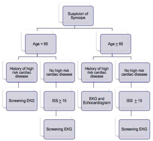
Is it possible to determine a subset of this population that may show a higher yield for this screening? Surgeons at Temple University in Philadelphia found that there was little utility in using carotid duplex studies. They did note that patients with a history of heart disease were more likely to have an abnormal EKG, and that an abnormal EKG predicted an abnormal echo. Overall, only patients with a history of significant cardiac comorbidity, older age, and higher ISS had findings requiring intervention.

Bottom line: Don’t just reflexively order a syncope workup when there is a question of this problem. Think about it first, because the majority of these studies are nonproductive. They are not needed routinely in trauma patients with “syncope” as a contributing factor.  Obtain a good cardiac history, and if indicated, order an EKG and go from there. See the practice guideline proposed by the Temple group below. And be sure to include the patients primary doctor in the loop!

References:

  1. Routine or protocol evaluation of trauma patients with suspected syncope is unnecessary. J Trauma 70(2):428-432, 2011.
  2. Syncope workup: Greater yield in select trauma population. Intl J Surg, accepted for publication June 27, 2017.