Thoughts On Traumatic Hematuria: Part 2

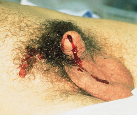
Yesterday, I discussed blood in the urine from a urethra. As I mentioned, there is typically not much from that particular injury. Today, I’ll dig into the three causes of real hematuria.

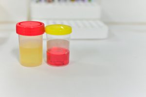
All of these tubes show gross hematuria except the one on the right.

  • Bladder injury. This can occur with either blunt or penetrating injury. The degree of hematuria is variable with stabs or gunshots, but tends to be much darker in blunt injury. This happens because the size of the bladder injury tends to be greater with blunt force. The bladder injury is not necessarily full-thickness with blunt trauma. It may just be some wall contusion and underlying mucosal injury. But frequently, with seat belt injury and/or A-P compression injuries to the pelvis (“open book”), the injury is full thickness.
    • Tip: If less than 50cc of very dark urine flow from the catheter upon insertion, it is likely that your patient has an intraperitoneal bladder rupture!
  • Ureteral injury. This injury is very rare. The most common mechanism is penetrating, but this structure is so small and deep that it seldom gets hit by naything. Patients with multiple lumbar transverse process fractures will occasionally have a small amount of hematuria, probably from a minor contusion. More often than not, the hematuria is microscopic, so we should never know about it.
  • Kidney injury. The most important fact regarding renal injury is that the degree of injury has no correlation with the amount of hematuria. The most devastating injury, a devascularized kidney, frequently has little if any gross hematuria. And conversely, a very minor contusion can produce very red urine.

So what about diagnosis? It’s easy! If you see gross hematuria, insert a foley catheter (if not already done) and order a CT of the abdomen/pelvis with contrast, as well as a CT cystogram. The latter must not be done using passive filling of the bladder with a clamped catheter. Contrast must be infused into the bladder under pressure to ensure a bladder injury can be identified.

CT scan is an excellent tool for defining injuries to kidney, ureter, and bladder, and will identify extravasation into specific places and allow grading. Specific management will be the topic of future posts.

Thoughts On Traumatic Hematuria: Part 1

I’ve seen a number of patients recently with bloody urine, and that is prompting me to provide some (written) clarity to others who need to manage this clinical problem. I’ll try to keep it organized!

There are two kinds of hematuria in trauma: blood that you can see with the naked eye, and…

Okay, so there’s only one. Trauma professionals do not care about microscopic hematuria. It does not change clinical management. Sure, your patient might have a renal contusion, but you won’t do anything about that. Or, he/she might have an infarcting kidney. And you can’t do anything about that. If you order a urinalysis, you might see a few RBCs. Don’t let this lead you down the path of looking for a source. You’ll end up ordering lots of tests and additional imaging, and generally will have nothing to show for it at the end. It’s not your job to spend good money on the very rare chance of finding something clinically significant.

Both of these specimens have blood in them. You can’t see it on the left, so don’t go looking for it with a microscope.

There are four sources of blood in the urine.

1. The first source does not generally cause hematuria, but can occasionally cause a few visible wisps of blood. That source is a urethral injury. The textbook teaching, and it’s good advice, is to look at the urethral meatus in your trauma patient, especially if you are contemplating insertion of a urinary catheter. If you see a few drops of blood, pause to consider. Sometimes, the blood is no longer visible, but might be present as a few well-placed drops on the patient’s underwear. So have a look at that, too, especially in patients with high risk injuries such as A-P compression pelvic fractures (think, lots of ramus fractures or pubic diastasis).

If you didn’t notice it and inserted the catheter anyway, you might see a few wisps of blood in the tubing as you place it. More often than not, this is just run of the mill irritation of the mucosa by the catheter, but always keep the possibility of an injury in mind.

Tomorrow, I’ll discuss the remaining three sources, and what to do about them.

Related posts:

Video: The Most Educational Trauma Surgeon In The World

Several readers asked me to dust off this video yet again. Enjoy this parody of the Dos Equis “Most Important Man In The World” commercials. I love poking fun at myself, and the slow motion shot on the helipad is hysterical.

This video was part of the Trauma Education: The Next Generation conference produced several years ago. Enjoy, and please comment or give it a thumbs up on YouTube!

Michael

YouTube player

How To: Needle Decompression Of The Chest

Here’s a quick, 3 ½ minute video for physicians and paramedics on how to decompress the chest when you suspect a tension pneumothorax.

The ATLS course now adds a consideration to use an alternative site. That location is the 5th intercostal space around the mid-axillary line. This has come about because shorter needles may not reach the pleural space when inserted under the clavicle in larger patients. The new spot is the typical location for placement of the inevitable chest tube that has to be inserted after needle decompression.

If you’ve got a few tips or tricks that you’d like to share on this procedure, please comment on the YouTube video.

YouTube player

Can Prehospital Providers Accurately Estimate Blood Loss? Part 2

I’ve previously written about the difficulties estimating how much blood is on the ground at the trauma scene. In general, EMS providers underestimated blood loss 87% of the time. The experience level of the medic was of no help, and the accuracy actually got worse with larger amounts of blood lost!

A group in Hong Kong developed a color coded chart (nomogram) to assist with estimation of blood loss at the scene. It translated the area of blood on a non-absorbent surface to the volume lost. A convenience study was designed to judge the accuracy that  could be achieved using the nomogram. Sixty one providers were selected, and estimated the size of four pools of blood, both before and after a 2 minute training session on the nomogram.

Here’s what it looks like:

Note the areas across the bottom. In addition to colored square areas, the orange block is a quick estimate of the size of a piece of paper (A4 size since they’re in Hong Kong!)

Here are the factoids:

  • The 61 subjects had an average of 3 years of experience
  • Four scenarios were presented to each: 180ml, 470ml, 940ml, and 1550ml. These did not correspond exactly to any of the color blocks.
  • Before nomogram use, underestimation of blood loss increased as the pool of blood was larger, similar to the previous study
  • There was a significant increase in accuracy for all 4 scenarios using the nomogram, and underestimation was significantly better for all but the 940ml group
  • Median percentage of error was 43% before nomogram training, vs only 23% after. This was highly significant.

Bottom line: This is a really cool idea, and can make estimation of field blood loss more accurate. All the medic needs to do is know the length of their shoe and the width of their hand in cm. They can then estimate the length and width of the pool of blood and refer to the chart . Extrapolation between colors is very simple, just look at the line. The only drawback I can see occurs when the blood is on an irregular or more absorbent surface (grass, inside of a car). 

Related posts:

Reference:  Improvement of blood loss volume estimation by paramedics using a pictorial nomogram: a developmental study. Injury article in press Oct 2017.