Trocar Chest Tubes Or Blunt Technique? Part 1

This is an old question: what is the best way to insert a chest tube? There are several techniques available to us:

  • Blunt dissection and insertion
  • Trocar with a blunt tip (plastic stylet)
  • Trocar with a sharp tip (metal stylet)
  • Seldinger technique for small tubes

Typically, when there are multiple ways to do a thing, then there is no clear choice as to which is better. It then becomes a personal choice, or one driven by the financial considerations of the equipment used, and demonstrates the need for a practice guideline.

There are very few good papers out there that critically compare any of these techniques. Today, I’ll review one cadaver study and tomorrow I’ll tackle one “best evidence” paper that attempt to answer it.

A group in Vienna, Austria performed a cadaver study comparing the use of the two types of trocar tubes:

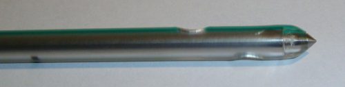
The top tube is the sharp trocar type, the bottom is the blunt trocar.

The study engaged twenty emergency medicine residents who had little, if any, experience placing chest tubes. Each placed 10 chest tubes (5 of each type) in fresh cadavers after undergoing a one-hour standardized lecture on anatomy, technique, and complications. The authors tabulated insertion times, as well as complication and success rate based on anatomic dissection.

Tube type was randomly assigned for each attempt by each resident. One blunt insertion and one sharp insertion were performed on opposite sides of a cadaver each month for the trainees. Over a period of 5 months, each resident performed 10 total insertions.

Here are the factoids:

  • Mean time to insertion for blunt vs sharp tips was the same, about 60 seconds
  • Insertion time declined by about 20 seconds by the final attempt at 5 months
  • Accurate placement occurred in 94% of blunt tip tubes vs 86% of sharp tip tubes
  • There were significantly more complications with the sharp tip (4 below diaphragm, 5 outside the thorax, 1 in the liver,  and 4 in the spleen) vs the blunt tip (2 below diaphragm, 2 extrathoracic, 2 in the liver, and 2 aborted due to damage to the tube)
  • BMI did not increase complications, but it did increase insertion time significantly

The authors concluded that there is a 6-14% complication rate that is operator related, and that the incidence of complications was increased with the use of a sharp tip tube. They warn against the use of these tubes.

Bottom line: This is certainly an interesting study. The insertion numbers are sort of reasonable, and the use of fresh cadavers is okay. They are not quite as realistic as real living people, but close. The biggest drawback was that they used chest tube newbies, most of whom had never inserted a tube. And they were placed in the unrealistic setting where they had to attend training and watch a video, then insert two tubes per month without coaching or supervision. This is not how we do it in the real world. 

I was impressed with what I consider the high number of complications. I don’t typically see that many, although I work at a blunt dissection institution. However, it does show that any trocar style tube is probably more like a weapon in inexperienced hands. So perhaps, even with supervision, both sharp and blunt trocar types should be avoided in the teaching setting. Sure, blunt dissection may take a bit longer, but the tube is also less likely to end up somewhere it shouldn’t be.

Tomorrow: Review of a “best evidence” review from New York.

Reference: Evaluation of performance of two different chest tubes with either a sharp or a blunt tip for thoracostomy in 100 human cadavers. Scand J Trauma Resus Emerg Med 20:10, 2012.

What Should We Call Them? NOAC vs DOAC

They are the bane of trauma professionals, the anticoagulants that cannot be easily or cheaply reversed. Yes, I’m talking about the direct thrombin inhibitors and the Factor Xa inhibitors. They were originally called NOACs, or novel oral anticoagulants since they were newer than the old standard, warfarin. But they’ve also been listed as DOACs (direct) or TSOACS (target-specific, just rolls off the tongue doesn’t it?).

Here’s a nice table I put together recently showing the all the common oral agents available. Click the image for a full-size, more readable image.

Dabigatran was the first of the newer oral agents, and it is the only direct thrombin inhibitor in the group. The rest are Factor Xa inhibitors. This is easy to remember if you look at their generic name. Each will contain “xaban.” Get it? Xa ban.

The daily cost of warfarin is about $7, while the daily cost of the others is around $16 per day. However, that does not take into account the cost of blood work to monitor INR in those taking warfarin, so it’s cost will be higher.

What I found most interesting was the cost of the reversal agents, if any. For warfarin it’s either a hit of 4-factor prothrombin complex concentrate or many bags of plasma. Praxbind for the DTI dabigatran appears to be a bargain! But look at the agent for the Xa inhibitors, Andexxa! Almost $50K per pop!

And what about the asterisk, you ask? That means that there is no literature available that shows that these expensive drugs are clinically effective! But they seem like they should work. Hmm.

Anyway, back to the nomenclature. NOACs or DOACs? Opinion is moving away from NOAC because it can be misinterpreted as “no anticoagulants.” The International Society on Thrombosis and Haemostasis polled their members, and the consensus opinion was that DOAC should be adopted for common use.  They add that the specific mechanism of action (direct thrombin vs Xa inhibitor) should be specified in addition to the DOAC acronym when clinically relevant.

Bottom line: DOAC wins! So hopefully we can all converge on using one common term for this group of drugs. Yet I still shudder when I have a head injured patient that tells me they are taking any of them!

Reference: Recommendation on the nomenclature for oral anticoagulants: communication from the SSC of the ISTH.  J Thrombosis Haemostasis 13(6):1154-1156, 2015.

Who’s Better At Invasive Procedures? Residents vs NP/PAs

With the implementation of resident work hour restrictions 15 years ago, resident participation in clinical care has declined. In order to make up for this loss of clinical manpower and expertise, many hospitals have added advanced clinical providers (ACPs, nurse practitioners and physician assistants). These ACPs are being given more and more advanced responsibilities, in all clinical settings. This includes performing invasive procedures on critically ill patients.

A study from Carolinas Medical Center in Charlotte NC compared complication rates for invasive procedures performed by ACPs vs residents in a Level I trauma center setting.

A one year retrospective study was carried out. Here are the factoids:

  • Residents were either surgery or emergency medicine PGY2s
    ACPs and residents underwent an orientation and animal- or simulation-based training in procedures
  • All procedures were supervised by an attending physician
  • Arterial lines, central venous lines, chest tubes, percutaneous endoscopic gastrostomy, tracheostomy, and broncho-alveolar lavage performances were studied
  • Residents performed 1020 procedures and had 21 complications (2%)
  • ACPs performed 555 procedures and had 11 complications (2%)
  • ICU and hospital length of stay, and mortality rates were no different between the groups

Bottom line: Resident and ACP performance of invasive procedures is comparable. As residents become less available for these procedures, ACPs can (and will) be hired to take their place. Although this is great news for hospitals that need manpower to assist their surgeons and emergency physicians, it should be another wakeup call for training programs and educators to show that resident education will continue to degrade.

Reference: Comparison of procedural complications between resident physicians and advanced clinical providers. J Trauma 77(1):143-147, 2014.

Trauma In Pregnancy 5: C-Section – When?

The perimortem C-section (PMCS) is a heroic procedure designed to salvage a viable fetus from a moribund mother. Interestingly, in some mothers, delivery of the fetus results in return of spontaneous circulation.

The traditional teaching is that PMCS should be started within 4-5 minutes of the mother’s circulatory arrest. The longer it is delayed, the (much) lower the likelihood that the fetus will survive.

The reality is that it takes several minutes to prepare for this procedure because it is done so infrequently in most trauma centers. Recent literature suggests the following management for pregnant patients in blunt traumatic arrest (BTA):

  • Cover the usual BTA bases, including securing the airway, obtaining access and rapidly infusing crystalloid, decompressing both sides of the chest, and assessing for an unstable pelvis
  • Assess for fetal viability. The fundus must measure at least 23 cm.
  • Assess for a shockable vs non-shockable rhythm. If shockable, do two cycles of CPR before beginning the PMCS. If non-shockable, move straight to this procedure.

Bottom line: Any time you receive a pregnant patient in blunt arrest, have someone open the C-section pack while you assess and try to improve the mother’s viability. As soon as you complete the three tasks above, start the procedure! You don’t need to wait 4 minutes!

Trauma In Pregnancy 4: Imaging

Everyone worries about imaging pregnant patients. As with most medical tests, it always boils down to risks vs benefits. What are the chances of causing mutations or cancers or a spontaneous abortion, and what is the risk of missing a critical injury? In general, reasonable studies involving a fetus at just about any point in gestation won’t cause major problems. At least as far as we know. What is not clear are the longer term, hard to measure effects. So the general philosophy should be to order just what you absolutely need, and shield the fetus during any studies other than of the abdomen/pelvis.

Now, to put these numbers into perspective, have a look at this list of delivered doses from common studies. The table above is listed in milliGrays, and this one is in milliSieverts. These are roughly comparable, except that the former is a measure of radiation dose absorbed, and the latter measures radiation delivered.

Bottom line: Think hard about the imaging you really need. If you generally do this for all patients, you probably won’t change your practice in pregnant women. Don’t worry about chest and pelvic x-rays. Shield the fetus for anything not involving the abdomen/pelvis. For major torso trauma, you probably will need CT of the chest/abdomen/pelvis. If so, do it right. Order with contrast so you don’t get substandard images that need to be repeated.