Trauma In Pregnancy 3: Tips & Tricks

Here is a list of practical tips for trauma professionals from prehospital and beyond:

  • Apply supplemental oxygen from the get-go, in the field if possible
  • Start an IV or IO and begin crystalloid infusion immediately
  • Bump the patient to her left. This is most easily done with the spine board in place. Just put a rolled up towel under the right side of the board.
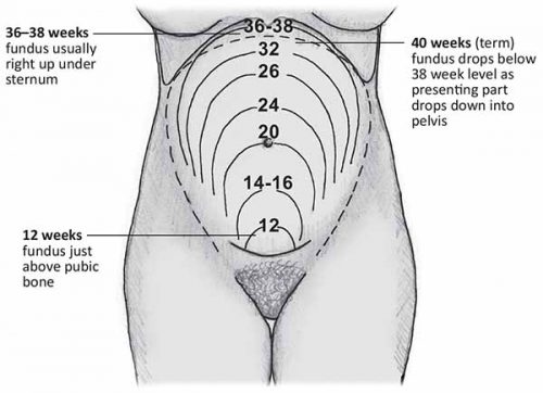
  • Quickly estimate how pregnant the patient is if she can’t tell you. Use the fundal height diagram below. Key measurements to remember:
    • Umbilicus = 20 weeks: entering the possible viability range if delivered emergently
    • Xiphoid = approaching full term

  • If the patient is 20 weeks+ pregnant, only transport to a trauma center (Level I or II).
  • At the hospital, reassess fundal height to more accurately estimate viability
  • If the fetus may be viable (23-24 weeks), call OB to the resuscitation area. But remember, they have nothing to offer until the trauma team secures the safety and stability of the mother. So don’t let them in the room until you have finished your entire evaluation.  Nothing they can do (e.g. monitor) is helpful during the acute phase of the resuscitation.
  • Once the mother is stabilized, assess fetal heart tones and apply monitors for fetal heart rate and maternal contractions.
  • Add coag studies to your lab panel. Fetal and placental injury can cause clotting problems.
  • Consider giving Rhogam in all major trauma cases if the mother is Rh negative
  • Obtain a KB test to look at the amount of fetal blood in the maternal circulation. Why do this if you are going to give Rhogam anyway? Answer: sometimes the amount of fetal blood leakage is greater than that covered by a single vial of Rhogam.
  • Plan your imaging intelligently. The next section covers this topic in more detail.
  • If needed, chest tubes should be inserted 1-2 intercostal spaces higher than usual.
  • The life-saving tetanus shot is safe in pregnant patients.


Trauma In Pregnancy 2: Predicting Outcome

The data on maternal outcome after trauma is mixed and somewhat confusing. Mortality after major trauma actually appears to be less. However, injury severity score (ISS) still correlates fairly well with overall mortality. But interestingly, other outcomes (complications) appear to be worse, even for relatively minor injuries. The reason behind this is not clear. Could it be a result of all of the physiologic changes noted above, hormonal factors, or something we don’t fully understand?

Fetal outcome is a function of the mechanism of injury (blunt vs penetrating), and extreme injury severity in the mother. Penetrating injury is uniformly devastating to the fetus, with 70% mortality for gunshots and 40% for stabs. Fetal death from blunt injury is primarily a function of placental abruption. About two thirds of blunt fetal deaths are due to abruption, with 50% of them due to car crashes. Maternal ISS does not correlate with fetal death, except in cases of very high scores. These women most likely experience anatomic and physiologic injuries that lead to fetal demise.

Tomorrow: Tips & Tricks

Reference: Trauma during pregnancy. OB Clinics of North America 40:47-57, 2013.

Trauma In Pregnancy 1: Introduction

Trauma in the pregnant patient is scary, but thankfully not very common. About 1 in 15 pregnant women sustain some type of significant injury. About half are from car crashes (most commonly if unrestrained), and 25% each are from assault (frequently domestic) and falls. Unfortunately, trauma is the leading cause of non-obstetric fetal death.

What makes this type of trauma problematic is two-fold, literally. First, you have two patients. And second, neither one has “normal” physiology. The mother has made numerous adaptations to accommodate the pregnancy, and the fetus is essentially a small parasite, totally dependent on her.

Let’s look at the impact of some of the more important maternal adaptations:

  • Total blood volume increases by about one liter. This would seem to be good, but since it is mainly dilutional with no real increase in total RBCs, and this is coupled with a lower mean blood pressure. Trauma professionals can easily underestimate blood loss.
  • Going into the third trimester, the uterus can compress the IVC when the mother is lying supine. but it is quite frequently forgotten. One of the first maneuvers when you suspect an advanced pregnancy is to bump the patient to her left. Do this even if the blood pressure is normal.
  • The pelvic veins are huge. Disruption from pelvic fractures or penetrating injury can lead to massive bleeding.
  • The stomach is probably full, and under pressure from below. This increases aspiration risk in women who have decreased mental status or need intubation.

And don’t forget the fetus:

  • The fetal/placental/uterine complex is one large, non-compressible unit with multiple shear planes within it. Blunt force will stress those planes, and may result in disruption of the uterus from pelvic veins (massive bleeding), or separation of the placenta from the uterus (abruption).
  • The fetus is totally dependent on the mother for survival, but the placenta will protect the mother first, shutting down fetal circulation if she becomes hypotensive or hypoxic.
  • The baby was designed to come into this world at full term. We have developed the technology to sustain life in smaller and more premature babies. The magic number of weeks keeps slowly decreasing, but preemie survival without complications is a challenge.

Tomorrow, we’ll move on and get to the fun stuff, predicting outcome after trauma in pregnancy.

Platelet Transfusion In Patients Taking Anti-Platelet Drugs

These days, trauma professionals see quite a few patients who take antiplatelet agents for cardiovascular comorbidities. These drugs can be problematic when the patients sustain injuries that result in bleeding in problematic areas like the cranial vault.

Aspirin and clopidogrel are the most common medications, and they irreversibly inhibit platelet aggregation.  All exposed platelets essentially quit working for the remainder of their 10-day lifespan. Platelet aggregation improves slowly over time after cessation of the drug as new platelets are added to the circulation from the bone marrow.

But what can you do if you are concerned that your patient is bleeding after injury because their platelets are not working? It seems logical that you would just transfuse some new platelets. But you should know by now that not everything that makes sense really works. A group in France designed a study to test this premise in patients taking either aspirin or clopidogrel. They performed a prospective, observational study on patients presenting with potentially life-threatening hemorrhage.

The authors used the Verify Now device to measure platelet response to the two drugs. Patients who had normal platelet function in the first place (not compliant or not a responder to the therapy) were excluded. All patients had initial platelet counts greater than 100K/ml. They underwent platelet transfusion for management of hemorrhagic shock, intracranial hemorrhage, or an emergent neurosurgical procedure.

Here are the factoids:

  • Only 25 patients were enrolled during the three year study; 13 were receiving only aspirin, 8 clopidogrel only, and 4 combined therapy
  • Average transfusions were 1-2 apheresis packs of platelets (6-12 units)
  • For aspirin patients, all showed significant platelet dysfunction before transfusion, and all but one showed recovery of function post-transfusion
  • For clopidogrel patients, platelet function remained impaired; the percent of inhibited platelets decreased but remained above the study threshold for “normal” of 20%

Bottom line: This is a very small study, but drives home the point that clopidogrel and its relatives may be problematic in bleeding patients. The active metabolites of this drug class are not well understood. But they are most likely still circulating in the blood in patients actively taking them, and deactivate new platelets as soon as they are transfused (assuming that the transfused platelets have good function in the first place). 

This issue requires further study so we can really tease out the actions of the drugs and their effect on transfused platelets. Until then, carefully consider whether platelet transfusion will be helpful in your bleeding patients, and if it is even worthwhile giving them or waiting for them to finish prior to going to the operating room.

Reference: Is platelet transfusion efficient to restore platelet reactivity in patients who are responders to aspirin and/or clopidogrel before emergency surgery? J Trauma 74(5):1367-1369, 2013.

What Would You Do? The Elderly Patient With Subdural Hematoma – Final Answer?

I’ve spent the last several posts reviewing the sparse data that we have on the impact of subdural hematoma management in elderly patients. With this information, I had hoped to arrive at some answers as to what to do when certain common patient presentations are encountered.

Unfortunately, the data is not very good, and is structured to raise false hopes. Overall, it looks like about 30-40% of selected patients die in the postoperative period. And the percentage of patients who are discharged at their pre-injury level of independence is in the low single digits. In fairness, one paper did show an improvement from severely disabled to moderately disabled or recovered, although the authors obfuscated how many actually made it to the good recovery group.

The biggest problem with all of the literature we have is that the patients were selected for surgery based on the opinion of the neurosurgeon. This means that many patients who they felt would do poorly with operation were excluded. It is extremely likely that the inclusion of these patients would have dragged down the already poor numbers that were reported. But in fairness, we might have also found a few surprise saves among those patients; I guess we’ll never know.

So let me give you my take on the scenarios that I presented so many days ago. Remember, these are my opinions and are not meant to be gospel. Other trauma professionals will need to interpret the information themselves and make their own decisions.

Scenario 1 – An elderly female falls and sustains a modest subdural hematoma with no shift and a normal exam. Follow your established practice guidelines unless some of the factors in the following scenarios are present.

Scenario 2 – Same as above, but the patient presents 8 hours after the fall. The clock started ticking when the fall occurred. Since your practice guideline recommends monitoring for 6 hours and then a followup CT of the head, the initial CT is the followup scan. The patient could then be discharged if there are no alarming findings on initial CT and the neuro exam is normal.

Scenario 3 – Same as scenario 1 but the patient has advanced dementia. These patients were generally excluded from the studies, and they are not expected to do well. Frankly, they will likely be much worse after an operation and will require an even higher level of post-discharge care (if they make it that far) and more involvement of family. It is critically important that the trauma professionals have a frank talk with the family to make sure they understand the overwhelming likelihood that their loved one will never be as good as they were before the injury. Surviving an operation does not mean going back to their usual living situation. The family absolutely needs this information to make the best choice for their loved one.

Scenario 4 – Same as scenario 1 but the patient has a well documented “do not actively resuscitate” order in place. The patient and their family need the same talk as above so they can appreciate all of the risks and the few, if any, benefits of surgery. Only then can they make an informed if they want to consider temporarily rescinding their DNAR order to allow surgery.

Scenario 5 – Same as scenario 1 but the patient is 95 years old. The data showed that patients in their 80s tended to do even more poorly than younger patients. There were very few nonagenarians in the literature, but it can be expected they would do even worse than the octos. They and their families need the same depressing talk so they can make the right decision.

Bottom line: Communication is key. And good data is even more key, although we have too little of it. For now, all we can do is paint a somewhat depressing picture of generally poor outcomes in highly selected patients. Hopefully we’ll have better data some day and can slice and dice things a little better. This may eventually allow us to offer surgery to those patients who will actually benefit from it the most.