Post-Embolization Syndrome In Trauma

A reader requested that I write about post-embolization syndrome. Not being an oncologist or oncologic surgeon, I honestly had never heard about this before, let alone in trauma care. So I figured I would read up and share. And fortunately it was easy; there’s all of one paper about it in the trauma literature.

Post-embolization syndrome is a constellation of symptoms including pain, fever, nausea, and ileus that occurs after angio-embolization of the liver or spleen. There are reports that it is a common occurrence (60-80%) in patients being treated for cancer, and there are a few papers describing it in patients with splenic aneurysm. But only one for trauma.

Children’s Hospital of Boston / Harvard Medical School retrospectively reviewed 12 years of their pediatric  trauma registry data. For every child with a spleen injury who underwent angio-embolization, they matched four others with the same grade of injury who did not. A total of 448 children with blunt splenic injury were identified, and (thankfully) only 11 underwent angio-embolization. Nine had ongoing bleeding despite resuscitation, and two had developed splenic pseudoaneursyms.

Here are the factoids:

  • More of the children who underwent embolization had extravasation seen initially and required more blood products.  They also had longer ICU (3 vs 1 day) and hospital stays (8 vs 5 days). Not surprising, as that is why they had the procedure.
  • 90% of embolized kids had an ileus vs 2% of those not embolized, and they took longer to resume regular diet (5 vs 2 days)
  • Respiratory rate and blood pressure were higher on days 3 and 4 in the embolized group, as was the temperature on day 5 (? see below)
  • Pain was higher on day 5 in the embolized group (? see below again)
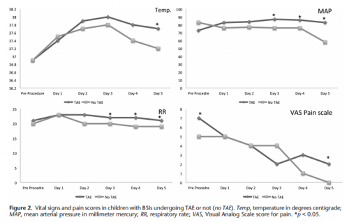
Bottom line: Sorry, but I’m not convinced. Yes, I have observed increased pain and temperature elevations in patients who have been embolized. Some have also had an ileus, but it’s difficult to say if that’s from the procedure or other injuries. And this very small series just doesn’t have enough power to convince me of any clinically significant differences in injured children.

Look at the results above. “Significant” differences were only identified on a few select days, but not on the same days across charts. And although the authors may have demonstrated statistical differences, are they clinically relevant? Is a respiratory rate of 22 different from 18? A temp of 37.8 vs 37.2? I don’t think so. And length of stay does not reveal anything because the time in the ICU or hospital is completely dependent on the whims of the surgeon.

I agree that post-embolization syndrome exists in cancer patients. But the findings in trauma patients are too nondescript. They just don’t stand out well enough on their own for me to consider them a real syndrome. As a trauma professional, be aware that your patient probably will experience more pain over the affected organ for a few days, and they will be slow to resume their diet. But other than supportive care and patience, nothing special need be done.

Related posts:

Reference: Transarterial embolization in children with blunt splenic injury
results in postembolization syndrome: A matched
case-control study. J Trauma 73(6):1558-1563, 2012.

Consequences Of Embolizing Renal Injuries

In my last post, I noted that nonoperative management is the norm for dealing with high grade renal injuries. One of the possible options, angioembolization, was relatively infrequently used at only 6% of the time.

For management of other organs like the spleen, there are several angioembolization options. Depending on the type and severity of injury, selective (partial) or nonselective (main splenic artery) embolization can be carried out. For the liver, only selective embolization can be used. But what about the kidney? 

Are there consequences of nonselective renal embolization? Or should we always strive for selective control? The urology group of the University of Tennessee – Knoxville published a series of papers on their experience using embolization in patients with the most severe injuries (Grade 5). They retrospectively examined just over 3 years of admissions with this injury. Numbers were very small (6 men, 3 women).

But they also published a second paper, extending the review dates to capture one more male patient. And they followed this group for 1.5 to 5 years (mean 2.5 years) to determine if any delayed complications surfaced.

Here are the factoids:

  • Seven patients underwent full, nonselective embolization, and the other three had “super selective” embolization
  • All patients had control of bleeding without surgical intervention
  • Followup CT imaging showed no persistent extravasation or expanding hematoma
  • No patient developed complications, such as a retroperitoneal abscess, prolonged fever, or hypertension while in the hospital or during short-term followup
  • Most patients showed a very small increase in serum creatinine (mean 0.04), but one patient increased from 1.1 to 1.7
  • On longer term followup, one patient, age 51, developed hypertension 10 months after his injury. It is not possible to determine whether he was one of the 20% of older adults who develop hypertension, or whether it was due to the procedure. it was well-controlled with a single antihypertensive med.
  • None developed altered renal function, stones, chronic pain, fistula, or pseudoaneurysm

Bottom line: Obviously, the data is very limited with only 10 patients. However, it is very interesting to note that the majority of these patients underwent nonselective embolization of the renal artery without any adverse event. The one case of hypertension occurred with nonselective embolization, although I have seen several case reports where this occurs with selective embolization as well.

It is now well-accepted that high-grade renal injury can and should be managed nonoperatively if the patient’s hemodynamic status is reasonable. I recommend a trip to interventional radiology if the patient has active extravasation or a high-grade (Grade 4 or 5) injury, as these patients are at risk for loss of the entire kidney otherwise. Selective embolization can be attempted first, but don’t be shy to take out the entire organ if need be. 

References: 

  • Percutaneous embolization for the management of Grade 5 renal trauma in hemodynamically unstable patients: initial experience. J Urology 181:1737-1741, 2008.
  • Intermediate-term follow-up of patients treated with percutaneous embolization for Grade 5 blunt renal trauma. J Trauma 69(2):468-470, 2010.

Contemporary Management Of Renal Injuries

A synopsis of contemporary management of renal injury was presented at the annual meeting of the American Association for the Surgery of Trauma last year. The Genito-Urinary Trauma Study Group (GUTS [groan!]) prospectively collected data on high-grade (grades 3-5) renal injuries from 14 Level I trauma centers over a 14 year period.

Here are some factoids from the article:

  • Expectant management (nonoperative or minimally invasive angio/stenting/drainage) was the norm, with 80% of these high-grade injuries dealt with in this manner
  • Only 6% of patients undergoing minimally invasive treatment underwent angioembolization
  • As expected, the higher the grade, the more likely the kidney would be removed (Grade 4 = 15%, Grade 5 = 62%)
  • Once operative management was performed, the nephrectomy rate escalated to 67%
  • Nephrectomy was more common in patients with penetrating trauma (60%)

Bottom line: Nonoperative management of renal injuries has long been the norm. This more recent review confirms it. Once the abdomen is opened, the chance of losing the entire kidney skyrockets. Expectant management (repeat exam and labs) is very common, and very successful. 

Angiography is an important adjunct, but was not used very commonly in this study. Perhaps the surgeons were concerned about complications from embolizing part or all of the kidney? I’ll discuss the consequences of this in my next post.

Reference: Contemporary management of high-grade renal trauma: Results from the American Association for the Surgery of Trauma Genitourinary Trauma study. J Trauma 84(3):418-425, 2018.

Ever Wonder Where The Golden Hour Came From?

Everywhere you turn in the trauma and EMS world, you run into the concept of the “golden hour.” Basically, it refers to the idea that it’s important to get an injured patient to definitive care promptly, or mortality begins to rise. It has been used to justify a lot of what we do in trauma care and trauma systems. But where did this come from? And is it true?

The BTLS course attributes the term to R Adams Cowley from the ShockTrauma Center in Baltimore. Unfortunately, no references are given. A biography of Cowley entitled Shock-Trauma names him the author of the term, basing it on dog research. No references were given.

A review of Cowley’s research reveals a few tidbits. A case series of patients implies that speed is good, but does not analyze time to definitive care. It does reference older work by other authors, but once again, no relationship between timing and outcome is evaluated.

A textbook edited by Cowley contains a reference to an article about “Cowley’s golden hour.” This article contains a statement that “patients are assumed to be dying and much of the golden hour has passed.” It goes on to state that the first 60 minutes after injury determines the patient’s mortality. It, in turn, refers to another of his earlier articles. This one states that “the first hour after injury will largely determine a critically injured person’s chance for survival.” No data or reference is given.

Bottom line: The concept of the “golden hour” has taken on a life of its own. Yes, it’s a good idea. And yes, there is some actual data to support it, although the quality is somewhat lacking. But this does point out the need to question everything, even some of our most deeply held beliefs. They are not always what they seem to be.

Reference: The Golden Hour: scientific fact or medical urban legend? Acad Emerg Med 8(7):758-760, 2001.

Complications Of Iliac Artery Embolization

The main cause of mortality in patients with severe pelvic fractures is major hemorrhage. Over the years, trauma professionals have developed and tested a number of maneuvers to reduce mortality in these patients. These include wrapping or fixing the pelvis, embolization, and more recently, pre-peritoneal packing and REBOA.

Pelvic wrap/fixation and embolization have been around for a long, long time. For both, it’s been long enough so that we should have a fairly decent appreciation of the complications. For pelvic binders, they principally involve the skin. But aside for the potential access site complications (bleeding, pseudoaneurysm), angiography has been thought to be relatively benign.

But as with any medical procedure, especially invasive ones, there are risks. A paper published five years ago retrospectively reviewed the 13 year experience with pelvic angiography at UC Davis. Study patients were matched with controls who underwent angiography for pelvic fracture but not embolization. Short-term (within 30 days) and long-term complications were assessed while in hospital and by telephone survey. Mean followup time was 18 months.

Here are the factoids:

  • There were no differences in complications attributable to embolization within 30 days of the procedure
  • There were 5 cases of short-term skin sloughing or necrosis in 55 patients, and 4 of 5 occurred in patients with nonselective embolization. However, this was not a statistically significant complication.
  • Long-term complications such as buttock claudication or skin ulceration, pain, and impotence were not significantly different in embolized vs non-embolized patients
  • There was a significantly increased incidence of buttock, perineal, or thigh paresthesias in the long-term

Bottom line: Angiography with embolization is a very valuable tool in the management of complication pelvic fractures. Remember that a number of complications have been described:

  • Skin sloughing or necrosis
  • Buttock claudication, pain, paresthesias
  • Skin ulceration
  • Impotence

Other than an increase in paresthesias in the long-term, there did not appear that there was any difference in patients undergoing angiography with and without embolization. Although the numbers were small (100 patients total), this is the best study we have to date. Just keep in mind that complications are possible, and question your patients about them when they present for their followup visits.

Reference: Evaluation of Short-term and Long-term Complications after Emergent Internal Iliac Artery Embolization in Patients with Pelvic Trauma. J Vascular Interventional Rad 19(6):840-847, 2008.