EMS: How Soon To Extricate The Pinned Patient?

This post was requested by one of my EMS colleagues who is the medical director of a rural EMS agency.

Maybe you watched the movie “Signs” by M. Night Shyamalan, starring Mel Gibson.  Gibson is a preacher whose wife was killed in a tragic accident. She was running and was pinned against a tree by a pickup truck. She is so badly injured that only the pressure of the truck against her is keeping her alive (and together, apparently). Gibson gets to have a few final words before being extricated (and killed).

Could this really happen? Shouldn’t entrapped people be extricated immediately, or do our prehospital providers need to wait until more advanced medical care is present at the scene?

Here’s the movie clip, if you are interested:

YouTube player

Obviously, you will find NO research on anything like this. The real question is, should EMS first responders (if not medically equipped and able) completely extricate an entrapped patient before paramedics or other trauma professionals with advanced skills are present? In other words, can you die just from being unentangled from the wreckage, like Mel Gibson’s wife?

The answer is, possibly. But it might not be for the reasons you think. Remember, this is Hollywood.

There are two killers upon release from entrapment. First, the mechanism by which the patient is pinned may be holding pressure on things that are or want to bleed. These include the pelvic bones, injuries to the torso, groins, and proximal extremities, and possibly even intra-abdominal hemorrhage sources. I’m discounting the chest because if there is enough pressure to tamponade bleeding, it will probably critically impair hemodynamics and ventilation to the point of killing your patient prior to extrication anyway.

The second factor is a crush injury, with release of a bolus of acidic, potassium laden blood from the crushed extremity upon release. This is probably quite rare, since it takes a significant amount of time for the un- or under-perfused extremity to build up enough of these substances to pose a threat. If the patient has been entrapped for less than 30-60 minutes, there is probably little danger to releasing them.

Bottom line: It is probably best to wait for ALS providers to arrive so IVs can be established and post-extrication resuscitation can be planned. This includes having fluid and/or blood products available in case critical bleeding starts once the pressure has been released. And don’t worry about reperfusion injury unless your patient has been trapped for quite a while.

Related posts:


A Sample Final Damage Control X-ray

Yesterday, I wrote about ways to reduce and hopefully eliminate retained foreign bodies (instruments, sponges) during damage control surgery. Today, I’ll provide a sample x-ray and some tips on how to use this tool most effectively.

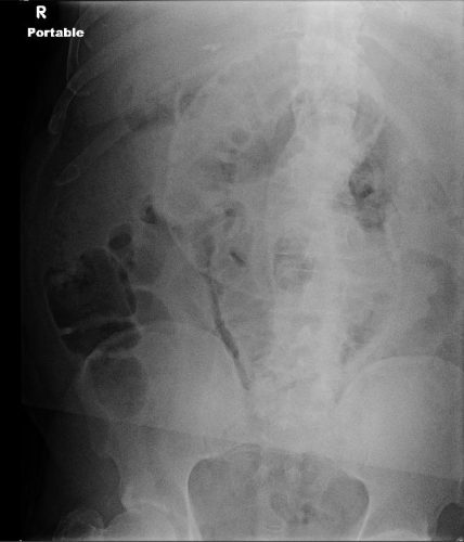
Here is an abdominal x-ray obtained just prior to closure of a patient who underwent damage control laparotomy. The OR record and surgeon from the initial operation documented that four sponges had been left in place for hemostasis.

dc-closure

Nothing retained, right?

Wrong! This image is not complete. This patient is larger than the x-ray plate used. The area under the diaphragms, the pelvis, and the entire left side of the peritoneal cavity have not been visualized.

Tips for imaging for damage control closure:

  • Always make sure the patient is on an x-ray OR table. It is so annoying (and potentially a sterility problem) to have to slide the plate under the patient!
  • Help the radiology tech to locate the desired imaging field using folds in the towels covering the body region. For example place the confluence of folds in the center of the towel in the exact place you want the center of the x-ray to be.
  • Remove all radiopaque objects from the x-ray field to reduce confusion when interpreting the image
  • Make sure the entire body cavity has been imaged! This may mean bracketing the area with several shots.
  • Read the image yourself! But if in doubt, or in patients with drains or other odd objects, call the radiologist to help you out.

Related posts:

The Final X-Ray In Damage Control Surgery

Damage control surgery for trauma is over 20 years old, yet we continue to find ways to refine it and make it better. Many lives have been saved over the years, but we’ve also discovered new questions. How soon should the patient go back for definitive closure? What is the optimal closure technique? What if it still won’t close?

One other troublesome issue surfaced as well. We discovered that it is entirely possible to leave things behind. Retained foreign bodies are the bane of any surgeon, and many, many systems are in place to avoid them. However, many of these processes are not possible in emergent trauma surgery. Preop instrument counts cannot be done. Handfuls of uncounted sponges may be packed into the wound.

I was only able to find one paper describing how often things are left behind in damage control surgery (see reference below), and it was uncommon in this single center study (3 cases out of about 2500 patients). However, it can be catastrophic, causing sepsis, physical damage to adjacent organs, and the risk of performing an additional operation in a sick trauma patient.

So what can we do to reduce the risk, hopefully to zero? Here are my  recommendations:

  • For busy centers that do frequent laparotomy or thoracotomy for trauma and have packs open and ready, pre-count all instruments and document it
  • Pre-count a set number of laparotomy pads into the packs
  • Use only items that are radiopaque or have a marker embedded in them. This includes surgical towels, too!
  • Implement a damage control closure x-ray policy. When the patient returns to OR and the surgeons are ready to begin the final closure, obtain an x-ray of the entire area that was operated upon. This must be performed and read before the closure is complete so that any identified retained objects can be removed.

Tomorrow, a sample damage control closure x-ray.

Related post:

Reference: Retained foreign bodies after emergent trauma surgery: incidence after 2526 cavitary explorations. Am Surg 73(10):1031-1034, 2007.

Tips For Surgeons: Abdominal Packing

One of the tenets of trauma surgery, handed down for generations, is that we should pack the abdomen to help manage major abdominal hemorrhage. “All four quadrants were packed” reads the typical operative note. But how exactly do you do that? Sounds easy, right?

sponges

Well, there are nuances not found in the surgery textbooks. Here are some practical tips for the trauma surgeon:

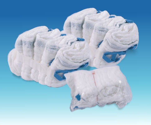
  • Prepare. Have your scrub nurse fluff up about 20 laparotomy pads in advance. The point of packing is two-fold: soak up blood, and stop bleeding. Fluffed up pads work better than the flat, rolled up pads shown above. And you will need them fast, so have a supply ready.
  • Do you really need to pack? Your patient is hypotensive, and you are convinced the abdomen is the source. You run to the OR, open it and… no blood. So don’t pack. It won’t slow down the (lack of) bleeding, but it is possible to cause serosal tears or worse. Just figure out where the bleeding is really coming from.
  • Be careful. Don’t just jam them in there. Carefully place pads over and under the liver. Carefully place a hand on the spleen and push toward the hilum so you can place pads between spleen and body wall. Try not to cause more damage than is already there.
  • Penetrating trauma: Pack where you know (or think) the penetrations are first. Basically, if it’s not bleeding there, don’t pack there.
  • Blunt trauma: Pack the upper quadrants first. This is where the money is, because the liver and spleen are the top culprits. Then pack the lower quadrants to soak up shed blood.
  • Once packed, check for successful control. If bleeding has stopped (or at least decreased significantly) stop and wait for anesthesia to catch up and continue your massive transfusion protocol. If bleeding continues, remove packs from the offending area and try to obtain definitive control. This is now the patient’s only chance, since you have now determined that you can’t stop the bleeding with packing.
  • Remove packs in the proper order. In blunt trauma, remove the lower quadrant packs first. They’re not doing anything and just take up valuable space. In penetrating trauma remove the packs in the area of the injury first.
  • Get an xray to confirm that all packs are out at the end of the case. Self explanatory. It’s easy to lose a few in the heat of the moment. I’ve seen two bundles (10 pads) left over the liver in one case decades ago!

On Monday, I’ll write about the importance of the final x-ray when the abdomen is closed.

Related post: