Trauma Activation Patients Staying Too Long In Your ED?

One of the long-held beliefs in trauma care relates to the so-called “golden hour.” Patients who receive definitive care promptly do better, we are told. In most trauma centers, the bulk of this early care takes place in the emergency department. However, for a variety of reasons, throughput in the ED can be slow. Could extended periods of time spent in the ED after patient arrival have an impact on survival?

Wake Forest looked at their experience with nearly 4,000 trauma activation patients who were not taken to the OR immediately and who stayed in the ED for up to 5 hours. They looked at the impact of ED dwell time on in-hospital mortality, length of stay and ventilator days.

Overall mortality was 7%, and the average time in the ED was 3 hours and 15 minutes. The investigators set a reasonable but arbitrary threshold of 2 hours to try to get trauma activation patients out of the ED. When they looked at their numbers, they found that mortality increased (7.8% vs 4.3%) and that hospital and ICU lengths of stay were longer in the longer ED stay group. Hospital mortality increased with each hour spent in the ED, and 8.3% of patients staying between 4 and 5 hours dying. ED length of stay was an independent predictor for mortality even after correcting for ISS, RTS and age. The most common cause of death was late complications from infection.

Why is this happening? Patients staying longer in the ED between 2 and 5 hours were more badly injured but not more physiologically abnormal. This suggests that diagnostic studies or consultations were being performed. The authors speculated that the knowledge, experience and protocols used in the inpatient trauma unit were not in place in the ED, contributing to this effect.

Bottom line: This is an interesting retrospective study. It reflects the experience of only one hospital and the results could reflect specific issues found only at Wake Forest. However, shorter ED times are generally better for other reasons as well (throughput, patient satisfaction, etc). I would encourage all trauma centers to examine the flow and delivery of care for major trauma patients in the ED and to attempt to streamline those processes so the patients can move on to the inpatient trauma areas or ICU as efficiently as possible.

Reference: Emergency department length of stay is an independent predictor of hospital mortality in trauma activation patients. J Trauma 70(6):1317-1325, 2011.

What The Heck Is It? Final Answer!

Yesterday, I posted the physical exam findings on this mystery object. A tiny puncture wound was present just to the right of the xiphoid on the lower chest wall, and a small sharp point was palpable.

Here’s how to deal with it:

Step 1. (Image 1) Don’t let it move or try to pull it out immediately! I didn’t want this thing to go in any further, or work it’s way out in case it was in the heart. I snapped a hemostat on the end of it for stabilization.

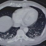
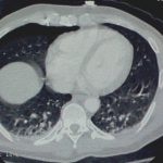
Step 2. (Images 2 and 3) Find out where it is exactly. You need to know what if any vital structures it may have pierced so you can plan for removal. In this stable patient, CT was the best option. If he had been unstable, it would have meant an immediate trip to the OR. Note how the object is within the chest wall, BUT it had penetrated at least as deep as the lung since a pneumothorax was present.

Step 3. (Image 4) Get it out! Off to the operating room for removal, just in case some unexpected bleeding or hemodynamic changes were to occur. After the patient was asleep, a chest tube was inserted. The object was a fine nail from a commercial nail gun mishap and pulled out easily with the hemostat. He was discharged two days later after the tube was pulled. 

Photo source: personal archive. Patient not treated at Regions Hospital

What The Heck Is It: Part 2

Last Friday I posted an xray that showed a long, thin metallic foreign body projecting from the right chest area toward the right side of the heart. What is it?

The patient declined to tell us what he had been doing. And looking at the xray, which is a 2D representation of a 3D object, it’s impossible to tell where the object is in the z axis. It could be in his back, in his clothes, or in his heart.

A complete physical exam showed a pinhole just to the right of the xiphoid process. By gently running a (gloved) finger across the hole, something thin and sharp was palpable. 

What should we do next? More images and the final answer tomorrow!

Results – Blunt Trauma Radiographic Imaging Protocol

In my previous post (click here to view) I discussed an imaging protocol that we developed and implemented last year. Today, I’ll detail what it has accomplished in our patients.

We looked at 229 patients who had their imaging performed according to the new protocol during a 3 month period and compared them to 215 patients who were imaged the previous year. Each scan administered to each body area (head, chest, abdomen/pelvis, c-spine, t-spine, l-spine, face, neck angio) were tabulated separately.

We found that the overall number of scans performed decreased significantly. We looked at our data and generated numbers per 100 patients. During the control period, we did 298 CT scans per 100 patients. This decreased to 271 during the study period. The number of head scans remained the same (82 per 100 patients during control, 85 per 100 during the study), as did the cervical spine scans (84 vs 86).

The biggest declines were seen in chest CT (53 per 100 control vs 33 per 100 study) and abdominal CT (57 vs 43).

We did see an increase in conventional xrays of the thoracic and lumbar spines to offset the absence of reformatted spine images that would have been generated from the chest and abdominal CT scans. We also noted small increases in CT of the head, cervical spine, and neck angio. This was likely due to better adherence to specific guidelines.

Bottom line: we believe that our work shows that careful adoption of well thought out guidelines can make a difference in practice and significantly decreases radiation exposure in our blunt trauma patients.

To read the post on this protocol, or to download it, click here.

Click here to download the Blunt Trauma Radiographic Imaging Protocol Worksheet