Tag Archives: stab

Nonoperative Management Of Abdominal Stab Wounds: The Practice Guideline!

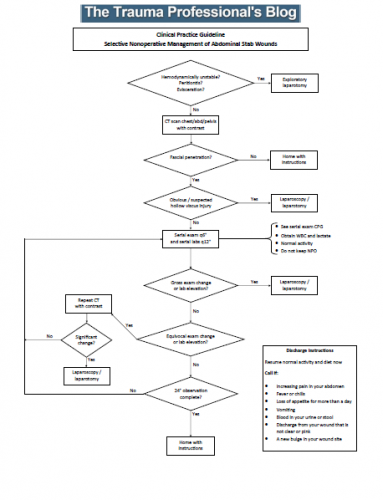
In my previous post, I reviewed a new paper that examined the appropriate amount of time that patients should be observed for nonoperative manage of an abdominal stab wound. Many of you know that I am a fanatic of properly crafted clinical practice guidelines (CPG). I decided to make a first pass at converting the LAC+USC group’s paper to something that will be helpful at the bedside.

This CPG incorporates the patient selection and timing information published in the paper. It breaks the process down into easily followed tasks, and fills in the blanks for shift to shift management. The CPG is displayed in an “if this, then do that” format. This firms up decision making and makes it easier for your trauma program to monitor compliance with it.

A note about CPGs: they generally cover about 90% of clinical cases. Obviously, they cannot provide guidance for certain rare combinations of circumstance. In that case, the trauma professional should do what they think is right for that situation. Most importantly, they should document this rationale in a progress note.

Here are answers to some of your questions in advance:

  • Patients should not be kept at bed rest. This is always bad.
  • There is no reason to keep the patient NPO. A very small percentage of patients actually fail. It makes no sense to starve everybody for the one or two patients that need to go to the OR each year. Anesthesiologists at trauma centers are very skilled at providing safe intubation in all patients. As you all know, every trauma activation patient coming into your trauma bay needing intubation has just finished a seven course meal!
  • Give your patient clear discharge instructions! They need to know what they can do, and what to look for if things eventually go awry.

And please leave comments and suggestions for improvements in the reply box below or by email to [email protected]. There are always ways to make CPGs even better! I have also included a Microsoft Publisher file so you can modify this guideline to better suit your trauma center.

In my next post, I’ll publish the serial abdominal observation CPG I mention in this one.

Resources:

  1. Download a pdf file of the guideline
  2. Download a Publisher file of the guideline


Nonoperative Management Of Abdominal Stabs: How Long Should We Watch?

Gunshots to the abdomen are a no-brainer that nearly always require operative exploration, but stab wounds are more challenging. They are low velocity, and injury only occurs in the pathway of the knife. It is more likely that inconsequential (or no) injuries occurred. Since exploratory laparotomy (or even laparoscopy) is not a benign procedure, trauma professionals frequently opt for selective nonoperative management (observation) in these cases.

What does observation mean? The patients are kept in the hospital for a set period of time, receive serial abdominal examinations, and get a few repeat lab tests. If the exam changes, the patient is taken to the OR to find out why. If it doesn’t, they are fed and sent home.

But what is the appropriate period of time to observe? One major concern is for the possible hollow viscus injury. Stomach and colon contain fluids that cause prompt peritonitis. But small intestinal content is rather innocuous, with neutral pH, normal concentration, and few bacteria. Peritonitis may not occur for days. Yet most centers send these patients home within 12 to 24 hours of injury.

What is the right answer? The trauma group at LAC+USC in Los Angeles performed a prospective, observational study to try to answer the question. They enrolled all patients with abdominal stab wounds presenting to their center over a three year period. They were generous (and correct) with their definition of the abdomen, including the thoracoabdominal portion up to the 5th intercostal spaces, and the pelvis.

Patients were excluded if they were hemodynamically unstable, or had an evisceration or obvious peritonitis. Most patients received a CT scan, and patients with suspected hollow viscus injury were excluded from the study and taken to the OR. All others were observed in a dedicated unit and were monitored for change in exam, need for blood transfusion, or other event which was recorded for the study.

Here are the factoids:

  • A total of 256 patients met study criteria: 77% had a single stab and 76% had a negative FAST exam
  • There were 46 patients who underwent immediate laparotomy for evisceration (59%), hemodynamic instability (33%), or peritonitis (24%)
  • 81% of patients underwent CT scan, and 13% were taken to OR based on the findings
  • This left 210 patients for nonoperative management
  • Of these, 71 had positive scans and all were due to solid organ injury. One patient failed at 32 hours due to increasing lactate and WBC, but the operation was nontherapeutic.
  • Another 14 patients had equivocal CT findings and two failed at 10 and 20 hours due to small bowel injury discovered by increasing lactate, WBC, change in exam, and air on a repeat CT
  • All 123 patients with negative scans passed nonoperative management
  • Median hospital length of stay was 3 with a range of 2-6 days

Since all of their patients who failed observation did so within 24 hours, the authors recommended a 24 hour observation period for all patients with stabs to the abdomen who did not meet their exclusion criteria.

Bottom line: Trauma professionals have needed a study like this for decades. Until now, we’ve been flying by the seat of our pants, with each surgeon making up his or her own magic number. This is a well done first attempt at defining what that number should be. 

Yes, there are some limitations to the study. The most important one that we don’t know the answer to is how many patients were successfully discharged that presented to another hospital with complications or failure. But this study provides a very reasonable estimate that helps us balance the cost (and patient inconvenience) of time in the hospital vs the dangers of a delayed diagnosis.

The only thing that remains is to design the practice guideline that incorporates the observation period, how often serial exams and labs should be obtained, and when the CT should be repeated.

Reference: Prospective evaluation of the selective nonoperative management of abdominal stab wounds: When is it safe to discharge? Journal of Trauma and Acute Care Surgery: November 2022 – Volume 93 – Issue 5 – p 639-643.

What Would You Do? Teensy Weensy Stab To The Abdomen – Part 2

Yesterday, I presented the case of a young man with a teensy weensy little stab to his abdomen, just above the umbilicus. There was a tiny bit of oddly colored fat that was visible in the wound. So now what should we do?

The first thing is to figure out what that bit of fat is. It doesn’t have the normal large pebbling and color of subcutaneous fat. Therefore, it must be a small piece of omentum protruding from the wound.

And what is the significance of that? This question has been addressed by papers with low numbers of subjects since the 1980s. It really depends on what country you are located in. Do you have readily available OR resources? Are there pressures to minimize hospital stays (US)?

One of the earliest papers originated from Parkland Hospital in Dallas TX. They reviewed 115 cases of omental evisceration over a 4 year period, and found that “serious” abdominal injuries were found in 75% of them. All went to laparotomy, and injuries to not one, but two organs were noted in about half of the positive cases. There was a 7% complication rate with negative laparotomy,

Contrast this with a study from Kingston, Jamaica where 66 patients with abdominal stabs and omental evisceration were treated. Of these, 14 were treated with observation because they had a normal abdominal exam. All were treated successfully without operation. But note the ratio here: 14/66 = 21%, which is the same as the negative laparotomy in the Parkland study (25%). So this study implies that if the patient can be watched and does not develop symptoms, the negative lap may be avoided.

Unfortunately, in many countries there are pressures to get people out of the hospital as soon as possible. Since small bowel content is relatively benign (at first), patients may not become symptomatic for several days. It would probably be difficult to convince your hospital to keep patients laying around for serial exams for days on end. Not to mention the logistical problems of doing good serial exams.

So most trauma professionals will be compelled to do something. And what should we do? Here are some possibilities. Pick your poison, and I’ll give you my choice tomorrow.

  • Local wound exploration
  • CT scan of the abdomen
  • Proceed to the operating room

As before, leave a comment to let me know what you would do. Or tweet it out!

References:

  1. Significance of omental evisceration in abdominal stab wounds. Am J Surg 152(6):670-673, 1986.
  2. Non-operative management of stab wounds to the abdomen with omental evisceration. J Royal Col Surg Edin 41(4):239-240, 1996.

Retained Foreign Objects After Penetrating Injury

A Chinese man was in the news a few years back after having a four inch knife blade removed from his head. It had been there for four years!  The knife blade broke off after he had been stabbed under the chin. Unfortunately, he was unaware that any part of the knife had been retained. It remained partly within the nasopharynx and the tip came to rest behind his left eye. His symptoms included headaches, stuffy nose and bad breath. The picture below shows the badly corroded blade in front of some of his radiographic images.

See the video at the bottom of this post for more details and images.

knife-in-head

What is the best way to deal with a problem like this? Here are some practical tips:

First, get in the habit of imaging any body part with a penetrating injury. Retained objects can be as simple as gravel or as complicated as the knife blade above. And remember, some patients who have been stabbed present with a simple laceration but don’t want to tell you how they got it. Image before you close it!

Next, don’t remove it. This is common knowledge, but innocent looking objects (pencils, nails) can penetrate arteries and keep them from bleeding while embedded. Unpleasant and sometimes fatal bleeding can ensue if pulled out.

If you do not have specialists versed in the body regions involved in the injury, transfer immediately with the object secured in place. For objects penetrating minimally complex areas like the extremities, surgeons may opt to carefully remove it in the emergency department, or may elect to do so in the operating room.

Injuries to complex areas should undergo high resolution CT scanning so that 3D reconstruction can be performed if needed. The surgical specialists can then plan the operative approach. This is dictated by the anatomy of the area(s) involved and the architecture of the object (think about hooks and barbs). For objects located near critical areas, an operative exposure must be selected that provides access to all portions of it, and allows for rapid vascular control if needed.

YouTube player

Penetrating Injuries to the Extremities

Simple penetrating injuries to the arms and legs are often over-treated with invasive testing and admission for observation. Frequently, these injuries can be rapidly evaluated and disposed of using physical examination skills alone.

Stabs and low velocity gunshots (no rifles or shotguns, please) should be thoroughly examined. This includes an examination of the entire, unclothed body. If this is not carried out, there is a risk that additional penetrating injuries may be missed.

For gunshots, look at the wounds and the estimated trajectory to try to demonstrate that the object stayed clear of neurovascular structures. This exam is imprecise, and must be accompanied by a full neurovascular exam and evaluation of the bones and joints. If there is any doubt regarding bony involvement, plain radiographs with entry markers should be performed. Any abnormal findings will require more in-depth evaluation and inpatient admission.

If the exam is negative but the trajectory is “in proximity” to a major vessel, an arterial pressure index (API) should be measured. This test involves the calculation of the ratio of the systolic pressure in the injured extremity to the contralateral uninjured extremity. It should not be confused with the ankle brachial index (ABI) which compares the systolic pressure in the ipsilateral uninjured arm  or leg.

The magic ratio is 0.9. If the API is less than this, there is some likelihood that a vascular injury is present. If the API is higher, there is virtually no chance of injury.

The final test that must be performed before discharge is a function test. If the injured extremity is too painful to use or walk on, the patient may need to be admitted for pain management and therapy. Patients managed in this way can avoid arteriography, CT angiography or admission and save thousands of dollars in hospital charges.

Reference: Journal Am Coll Surgeons 2009;209:740-5.