Tag Archives: chest

Detecting Rib Fractures In The Elderly

It’s well known that our elders do less well than younger folks after injury. The number of complications is higher, there tends to be more loss of independence during recovery, and mortality is increased. This is not only true of high energy trauma like car crashes, but also much lower energy events such as a fall from standing.

Rib fractures are common after falls in the elderly and contribute to significant morbidity if not treated adequately. Traditionally, they are identified through a combination of physical exam and chest x-ray. Unfortunately, only half of rib fractures are visible on x-ray. It falls to the physical exam to detect the rest.

A group at Beth Israel Hospital in Boston explored the utility of using chest CT in an attempt to determine if this would result in more appropriate and cost-efficient care in the elderly. They performed a retrospective study of 3 years of their own data on patients aged 65 or more presenting after a mechanical fall and receiving a rib fracture diagnosis. Imaging was ordered at the discretion of the physician. A total of 330 patients were elderly, fell, and had both chest x-ray and chest CT obtained. This was a very elderly group, with a mean age of 84 years!

Here are the factoids:

  • Rib fractures were seen on chest x-ray in 40 patients (12%) and on CT in an additional 56 ; 234 patients had no fractures on either
  • When fractures were seen on both studies, CT identified a median of 2 more fractures than chest x-ray
  • Patients with fractures not seen on chest x-ray were admitted significantly more often than those without fractures (91% vs 78%)
  • Mortality, admission to ICU, ICU length of stay, and hospital length of stay were not different if fractures were seen only on CT
  • CT scan identified new issues or clarified diagnoses suggested by chest x-ray in 14 cases, including one malignancy
  • Rib detail images were obtained in 13 patients and proved to be better than chest x-ray, but not quite as good as CT scan

Conclusion: use of CT for rib fracture diagnosis resulted in a few more admissions, but no change in hospital resource utilization, complications, or mortality.

Bottom line: Hmm…, read the paper closely. The authors conclude that more patients with CT-only identified rib fractures are admitted. But compared to what? Unfortunately, patients without rib fractures on CT. What about comparing to patients who had fractures seen on chest x-ray too? If that number is the same, then of what additional use is CT? Identifying a few incidentalomas?

Given that there is no change in the usual outcome measures listed here, it doesn’t seem like there is any additional benefit to adding CT. And I can see a lot of downsides: cost, radiation, and possible exposure to IV contrast. In my mind, there is still nothing that beats a good physical exam and a chest x-ray. Skip the CT scan. And don’t even think about ordering rib detail images! That’s so 1990s. And even if no rib fractures are seen on imaging, physical exam is the prime determinant for admitting your patient for aggressive pain management and pulmonary toilet.

Reference: Chest CT imaging utility for radiographically occult rib fractures in elderly fall-injured patients. J Trauma 86(5):838-843, 2019.

Detecting Rib Fractures In The Elderly

It’s well known that our elders do less well than younger folks after injury. The number of complications is higher, there tends to be more loss of independence during recovery, and mortality is increased. This is not only true of high energy trauma like car crashes, but also much lower energy events such as a fall from standing.

Rib fractures are common after falls in the elderly and contribute to significant morbidity if not treated adequately. Traditionally, they are identified through a combination of physical exam and chest x-ray. Unfortunately, only half of rib fractures are visible on x-ray. It falls to the physical exam to detect the rest.

A group at Beth Israel Hospital in Boston explored the utility of using chest CT in an attempt to determine if this would result in more appropriate and cost-efficient care in the elderly. They performed a retrospective study of 3 years of their own data on patients aged 65 or more presenting after a mechanical fall and receiving a rib fracture diagnosis. Imaging was ordered at the discretion of the physician. A total of 330 patients were elderly, fell, and had both chest x-ray and chest CT obtained. This was a very elderly group, with a mean age of 84 years!

Here are the factoids:

  • Rib fractures were seen on chest x-ray in 40 patients (12%) and on CT in an additional 56 ; 234 patients had no fractures on either
  • When fractures were seen on both studies, CT identified a median of 2 more fractures than chest x-ray
  • Patients with fractures not seen on chest x-ray were admitted significantly more often than those without fractures (91% vs 78%)
  • Mortality, admission to ICU, ICU length of stay, and hospital length of stay were not different if fractures were seen only on CT
  • CT scan identified new issues or clarified diagnoses suggested by chest x-ray in 14 cases, including one malignancy
  • Rib detail images were obtained in 13 patients and proved to be better than chest x-ray, but not quite as good as CT scan

Conclusion: use of CT for rib fracture diagnosis resulted in a few more admissions, but no change in hospital resource utilization, complications, or mortality.

Bottom line: Hmm…, read the paper closely. The authors conclude that more patients with CT-only identified rib fractures are admitted. But compared to what? Unfortunately, patients without rib fractures on CT. What about comparing to patients who had fractures seen on chest x-ray too? If that number is the same, then of what additional use is CT? Identifying a few incidentalomas?

Given that there is no change in the usual outcome measures listed here, it doesn’t seem like there is any additional benefit to adding CT. And I can see a lot of downsides: cost, radiation, and possible exposure to IV contrast. In my mind, there is still nothing that beats a good physical exam and a chest x-ray. Skip the CT scan. And don’t even think about ordering rib detail images! That’s so 1990s. And even if no rib fractures are seen on imaging, physical exam is the prime determinant for admitting your patient for aggressive pain management and pulmonary toilet.

Reference: Chest CT imaging utility for radiographically occult rib fractures in elderly fall-injured patients. J Trauma ePub ahead of print, Jan 23, 2019.

Don’t Repeat Trauma Activation X-rays!!

You are in the middle of a fast-paced trauma activation. The patient is awake, and mostly cooperative. The x-ray plate is under the patient and everyone stands back as the tech gets ready to fire the x-ray machine. At that very moment, your patient reaches up and places his hand on his chest. Or one of the nurses reaches over to check an IV site.

The x-ray tech swears, and offers to re-shoot the image. What do you do? Is it really ruined? They have an extra plate in hand and are ready to slide it under the patient bed.

The decision tree on this one is very simple. There are two factors in play: what do you need to see, and how hard is it to see? The natural reaction is to discard the original image and immediately get a new one. It’s so easy! Plus, the techs will take heat from the radiologist because of the suboptimal image. But take a look at this example of a “ruined” chest xray.

It’s just the patient’s hand! You can still see everything that you really need to.

Bottom line: You are looking for 2 main things on the chest x-ray: big air and big blood. Only those will change your management in the trauma bay. And they are very easy to see. Couple that with the fact that an arm overlying the image does not add a lot of “noise” to the image. So look at the processed image first. 99% of the time, you can see what you need, and will almost never have to repeat. [Hint: the same holds true for the pelvic x-ray, too. You are mainly looking for significant bony displacements, which are also easy to see.]

Related posts:

The Right Way to Treat Tension Pneumothorax

Tension pneumothorax is an uncommon but potentially lethal manifestation of chest injury. An injury to the lung occurs that creates a one-way valve effect, allowing a small amount of air to escape with every breath. Eventually the volume becomes so large as to cause the lung and mediastinum to push toward the other side, with profound hypotension and cardiovascular collapse.

The classic clinical findings are:

  • Hypotension
  • Decreased or absent breath sounds on the affected side
  • Hyperresonance to percussion
  • Shift of the trachea away from the affected side
  • Distended neck veins

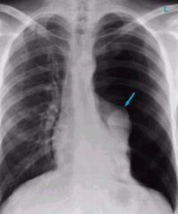
You should never diagnose a tension pneumothorax with a chest xray or CT scan, because the diagnosis is a clinical one and the patient may die while these procedures are carried out. Having said that, here’s one:

image

The arrow points to the completely collapsed lung. Note the trachea bowing to the right.

As soon as the diagnosis is made, the right thing to do is to “needle the chest.” A large bore angiocath should be placed in the second intercostal space, mid-clavicular line, sliding right over the top of the third rib. The needle should then be removed, leaving the catheter.

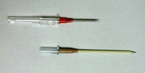
The traditional large bore needle is 14 gauge, but they tend to be short and flimsy. They may not penetrate the pleura in an obese patient, and will probably kink off rapidly. Order the largest, longest angiocath possible and stock them in your trauma resuscitation rooms.

image

The top catheter in this photo is a 14 gauge 1.25 inch model. The bottom (preferred at Regions) is a 10 gauge 3 inch unit. Big difference! 

The final tip to treating a tension pneumothorax is that a chest tube must be placed immediately after inserting the needle. If the patient is on a ventilator, the positive pressure will slowly expand the lung. But if they are breathing spontaneously, the needle will change the tension pneumothorax into a simple open pneumothorax. Patients with other cardiovascular problems will not tolerate this for long and may need to be intubated if you dawdle.

Related post:

Answer: What The Heck Video! Part 2

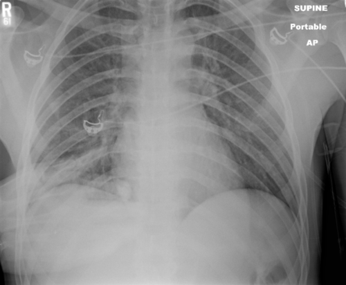
This patient was involved in a motor vehicle crash with significant chest trauma. They’ve been intubated and are oxygenating and ventilating well. What to do next?

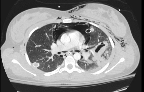
First, the endotracheal tube was a bit deep, which can create its own problems. It was pulled back a few centimeters. Since the patient was hemodynamically stable, a CT angio of the chest would be very helpful to try to figure out the pathology. Here’s a representative slice from the scan.

There are a few striking findings here:

  • Extensive subcutaneous emphysema
  • Large pneummediastinum around the heart
  • Significant injury to the left lung (note the pneumatocele, an air filled collection)
  • Atelectasis of the left lung despite repositioning of the ET tube

The combination of of the above is highly suggestive of a large airway injury. Since the entire lung was affected, it is most likely a mainstem bronchus injury. Usually, these are accompanied by a large air leak from the chest tube, but not in this case.

This prompted the bronchoscopy shown two days ago. The image is oriented such that the left mainstem bronchus was on the right side of the video. A bronchial tear is visible on the lateral aspect, just before the takeoff of the upper lobe bronchus. You can get the impression of a beating heart beating somewhere nearby. And when the camera pops through the laceration, you can actually see the thoracic aortic coursing away toward the diaphragm!

Related post: