Galleries

The Future of the Medical Journal – Part 2

After posting yesterday, I was informed that LWW was not the first to enter the small screen arena. The British Medical Journal (BMJ) released content for the iPad platform beginning in December 2011. This journal uses the built-in Newsstand app on the iPad or iPhone.

On first look, it appears to be more robust than the LWW app for Neurosurgery. The BMJ allows access to other related content streams, such as their news, blogs, podcasts and video. It is a bit more interactive and does allow searching and bookmarking.

Bottom line: I’m sure someone will step forward with yet another earlier entry into the smartphone/tablet arena. Regardless, it’s all good. Either Newsstand or independent app works, although the Newsstand offers some pre-built functionality for developers that might make it a bit easier to create. However, the Newsstand is also Apple-specific, and won’t work for all the people with Android or Windows based phones and tablets who will be screaming for this content.

Reference: search for “BMJ” in the Apple App Store

The Future of the Medical Journal

Journals have been printed on trees since forever. Within the past 10 years, there has been a shift toward also making this content available on the publishers’ web sites. Now, Lippincott Williams & Wilkins (LWW), publisher of the journal Neurosurgery (and many others) has taken an important step forward.

Beginning with the current issue (March 2012), this journal is now available within an iPad app. One of the problems with the traditional journal was that you had to go to the library to read it. And medical libraries can’t carry every journal, so if the one of interest was not in the collection, you had to wait a few days for the librarian to get a copy.

With the onset of the internet age, journals began to appear online. Most major publishers had a web presence and made their journals available online. However, most provided full text content for free only to their subscribers. At best, a non-subscriber would get the abstract, or an occasional free article. Biomedical libraries typically subscribed to services where hundreds of journals were available as full text to medical staff members, residents and students. The downside was, and still is, that you need access to a desktop to comfortably consume the articles. The web sites were just not that friendly for the small screen.

Finally, LWW has made a concerted effort to provide this content in tablet format. More and more trauma professionals are carrying these devices and using them in their practices. This brings the content closer to the patient and provides it in a very consumable format. It also allows the publisher to add dynamic digital content (audio and video) to the material.

I’ve been using this app for about a day and am impressed. Here are some key features:

  • Portability is excellent. A continuous internet connection is not needed. The entire journal issue is downloaded and added to your library, and you are only limited by the amount of storage on your iPad.
  • Extra digital content is routine. And since it is already downloaded with the issue, there is no waiting for it to load.
  • Articles have the same look and feel as the journal and can be panned or zoomed for readability.
  • Content sharing is possible (somewhat). Each article allows the user to share on Twitter or Facebook. The article can also be emailed to others. However, only a link to the article is provided, and if you are not a journal subscriber you’ll either have to pay up or use your medical library account to get full text when the link is opened.

What doesn’t it do? There is currently no ability to search through an article or issue. And it would be nice to interact with one of the authors via an email link to ask questions or make suggestions. Finally, Apple is not the only tablet maker out there. Publishers will need to make sure apps are available on the Android platform as well.

Bottom line: This is a great first-try at moving journals onto a mobile platform. I expect that LWW will begin to roll out this format for all their journals since they’ve now figured out how to do it. And expect all the other publishers to jump on the bandwagon as well. Journals are eventually going to go the way of DVD movies, and we’ll end up streaming our professional content from some company with a big red logo.

Reference: Search for “Neurosurgery” in the Apple App Store.

Pelvic Fractures: OR vs Angio In The Unstable Patient

One of the cardinal rules of trauma care is that hemodynamically unstable patients can only go the the operating room from the ED. No trips to CT, xray, etc. Trauma professionals occasionally try to make exceptions to the rule, but it usually doesn’t work out.

Well, what about the patient with severe pelvic fractures who is or becomes unstable? Pelvic fracture bleeding is not always easy or even possible to control in the OR, and angiography offers a way to identify and stop the bleeding, right?

The trauma group at Ryder in Miami did a lengthy (13 year) retrospective review of their experience with these patients. They looked at every patient who underwent angiography, then identified the subset that went to the OR followed by angiography. There were 134 angio patients and 49 OR to angio patients on whom they based their analysis. Obviously, there is plenty of opportunity for bias in this study, and many of the study patients identified had to be excluded due to incomplete records.

Patients who went to the OR first tended to have similar injury severity but were sicker than the angio alone group. Crystalloid and blood resuscitation volumes were significantly higher in the OR group as well. Most of these patients underwent a laparotomy, and 64% had active intra-abdominal bleeding. None died in OR, and most were left with a damage control abdominal closure.

In the angio group, there were really 2 subsets: angio alone, and angio followed by OR. Mortality in the angio alone group was similar to the OR-angio group. But deaths skyrocketed in those who went from angio to OR (67% vs 20%). This is likely due to them failing angiographic management of bleeding. Three patients died in the angio suite.

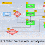
Bottom line: There’s a lot of data in this paper, and some of the results can be explained by selection bias. However, they appear to support algorithms released by EAST and the WTA (see diagram above). In general, a trauma patient with severe pelvic fractures and hemodynamic instability needs to go to OR to identify and treat any source of intra-abdominal bleeding. If pelvic bleeding remains a problem, preperitoneal packing may be considered, followed by a trip to angio at that point. The rule that unstable patients should only go to OR (or an ambulance bound for a trauma center if there is no OR) still holds!

Reference: Operating room or angiography suite for hemodynamically unstable pelvic fractures? J Trauma 72(2):364-372, 2012.

Quiz: There is just one extremely rare reason that I know of to move to CT with a hemodynamically unstable trauma patient. Leave a comment with your guess.

Temporal Bone Fracture

The temporal bone is a very complicated and important piece of bone located at the base of the skull. It houses a number of structures vital to hearing and balance, and is a conduit for several important nerves. Most fractures are due to blunt trauma, and although several classification systems exist, the majority are irregularly shaped and don’t lend themselves to easy categorization.

Clinically, a t-bone fracture should be suspected anytime that blood is seen in the ear canal or behind the eardrum after blunt head trauma. Patients may complain of dizziness or hearing loss. Examination may demonstrate some nystagmus in some cases. Most of the time, the diagnosis will not be made until a head CT is performed. The diagram above shows why this fracture can cause hearing problems (middle ear) or dizziness (inner ear).

There is usually no specific surgical intervention needed for this fracture. However, two important functional exams must be completed once diagnosed: facial nerve function and hearing. The facial nerve exam should document whether only portions of the nerve are affected vs all branches, and whether there is at least partial function. In head injured patients that can’t follow commands, even a grimace can demonstrate some function.

The ideal hearing test is carried out with a tuning fork. However, this is not usually convenient in acute trauma patients. Whispering and rubbing fingers near the patient’s ear can be used as a quick and dirty test. Awake patients will be able to tell you if their hearing is decreased.

Bottom line: Suspect temporal bone injuries in patients with any signs of basilar skull fracture. If the diagnosis is suspected or confirmed by CT scan, document the best facial nerve and hearing exam that you can. Contact your facial surgeon once the diagnosis is confirmed, and call them urgently if there is any loss of facial nerve function. Thin cut CT scans of the temporal bone are generally not necessary, and should not be ordered automatically unless the facial surgeon needs it for a specific reason.

Pneumothorax And Oxygen: The Final Post!

Okay, this is the last time I’m going to write about this. Hopefully I can provide the final nail in the coffin for this idea. Previously, the oldest paper I could find that was cited as a reason to use high inspired oxygen to treat pneumothorax was from 1983. I found what I think is the earliest (and the last that I will discuss) from 1971!

Twelve patients were retrospectively reviewed who recovered without intervention from a spontaneous pneumothorax. Another 10 were monitored prospectively with the same condition, but were given “high concentration oxygen” (??) by mask from 9 to 38 hours at a time. During intervening periods, the patients breathed room air. Daily chest xrays were obtained, and here is the cool part:

The inner edge of the chest wall and the outer edge of the lung were traced on transparent paper. This was then superimposed on graduated graph paper and the area corresponding to the pneumothorax cavity was measured. The rate of absorption was expressed in cm2/24 hrs.

Need I say more? The authors did show graphically that the apparent rate of absorption tripled in the treated patients, from about 5cm2/day to about 15cm2/day, and was higher in patients with a larger pneumothorax. The problem here is the same as before: chest xray does not allow volumetric estimates, so any results relying on them are suspect. At least it’s not a rabbit study.

Bottom line: There’s just no convincing data to support this practice, so let’s stop using it. Simple physics suggests that this should work, but the effect is just not clinically significant enough to offset the possibility of mishaps from an inpatient admission for oxygen therapy (see yesterday’s post). As I mentioned yesterday, look at the clinical status of your patient. If they have any detectable blood in their chest, they’ll probably need drainage. If not, and if they feel normal, discharge and follow up with a repeat xray in a week. The pneumo will probably be gone. If they do have some compromise, then insert the smallest tube you can. If done properly and a one-way valve can be used, the patient may still be managed as an outpatient.

Related posts:

Reference: Oxygen therapy for spontaneous pneumothorax. Br Med J 4:86-88, 9 Oct 1971.