Galleries

Novel Hip Reduction Technique: The Captain Morgan

I wrote about posterior hip dislocation and how to reduce it using the “standard” technique about 9 months ago. Emergency physicians and orthopedic surgeons at UCSF-Fresno just published their experience with a reduction technique called the Captain Morgan.

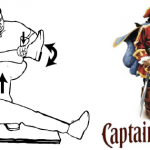
Named after the pose of the trademark pirate for Captain Morgan rum, this technique simplifies the task of pulling the hip back into position. One of the disadvantages of the standard technique is that it takes a fair amount of strength (and patient sedation) to reduce the hip. If the physician is small or the patient is big, the technique may fail.

In the Captain Morgan technique, the patient is left in their usual supine position and the pelvis is fixed to the table using a strap (call your OR to find one). The dislocated hip and the knee are both flexed to 90 degrees. The physician places their foot on the table with their knee behind the patient’s knee. Gentle downward force is placed on the patient’s ankle to keep the knee in flexion, and the physician then pushes down with their own foot, raising their calf. Gentle rotation of the patient’s hip while applying this upward traction behind the patient’s knee usually results in reduction.

Some orthopedic surgeons use a similar technique, but apply downward force on the patient’s ankle, using the leverage across their own knee to develop the reduction force needed. The Captain Morgan technique use the upward lift from their own leg to develop the reduction force. This may be gentler on the patient’s knee.

The authors report a series of 13 reductions, and all but one were successful. The failure occurred due to an intra-articular fragment, and that hip had to be reduced in the operating room. 

I’m interested in hearing comments from anyone who has used this technique (or the leverage one). And does anyone have any other techniques that have worked for them?

Related post:

Reference: The Captain Morgan technique for the reduction of the dislocated hip. Ann Emerg Med (in press) dol:1016/j.annemergmed.2011.07.010, 2011.

Thanks to Sam Stellpflug MD at Regions Hospital for bringing this article to my attention.

Weird Trauma: Pruning Shears to the Head and Neck

This case made the national news yesterday, and I wanted to make a few comments on the ideal management of this type of injury.

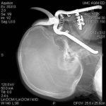
An 86 year old Arizona man was trimming plants in his back yard and fell on his pruning shears. One of the handle grips pushed into his orbit and through his pharynx into his neck. How do you think through something like this?

First, always check vital signs. If the patient is hypotensive, they must go to the operating room. Even if vital signs are stable, ongoing bleeding necessitates an operation before anything else.

If vital signs are stable, then a road map showing vial structures is essential. The patient should be taken to CT so the exact position of the object can be determined. Any involved structures (carotid artery, esophagus) can be identified and a proper plan can be developed. 

Then and only then can a stable patient be taken to the OR. Appropriate incisions should be placed so that key portions of the foreign object can be viewed as they are removed. In this case, incisions were made under his lip and into the maxillary sinus wall to monitor the removal process. The carotid artery had been cleared by CT. Once removed, any remaining bleeding can be addressed. 

A final point: any problem like this that has the potential to involve the airway requires that a skilled anesthesiologist be present with appropriate airway management equipment, and the surgeon needs to have all equipment ready to place a tracheostomy on short notice.

This patient did well after removal and was treated with about 3 weeks of antibiotics for his sinus injuries. His inferior orbital wall was rebuilt, and overall he did well postoperatively. He is seriously reconsidering doing any gardening again.

Return To Work After Severe Trauma

One of the most important goals after injury is return to work or school. There are some studies available that look at return to work/school status as a function of injury severity, demographic and insurance status. However, long-term studies are rare.

A Norwegian group followed a small population of injured patients very closely for five years, looking at the actual trajectory of return. They also tried to determine the specific factors that predicted return to work. The initial group numbered 101 people, but slowly decreased to 75 due to dropouts, nonresponders, and one patient who retired while receiving disability benefits.

The average age was 39 and ISS was 29. About 60% had a lower level of education and blue collar jobs. There were 28 patients with severe head injury, 12 with moderate head injury, 18 spinal cord injuries and 3 amputees among the group.

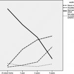
At the end of 5 years, only 49% had returned to work (see chart). 23% were on full disability and 9% on partial disability. Of greatest interest, there was only a small increase in return to work after 2 years. The best predictors of return to work were higher education level, good physical health and function (no surprise), and type of coping strategy. Time spent in rehab was also a factor.

Bottom line: Rehab that aims toward return to work is a major factor in getting better after major injury. However, an additional focus on coping and other psychological factors is important. Most people who will be capable of returning to work or school will do so by the two year mark.

Reference: Returning to work after severe multiple injuries: multidimensional functioning and the trajectory from injury to work at 5 years. J Trauma 71(2):425-434, 2011.