Tag Archives: what the heck?

What The Heck? Final Answer

So what is going on with your dyspneic patient after partial ejection from their car? They are not responding to your interventions as they should How is he immune to chest tube placement?

Well, you could intubate the patient. But the x-ray will be available momentarily, and may guide you along a different path. It might even obviate the need for intubation. So while you are getting your meds ready (just in case), this image comes up:

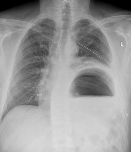
The chest tube is in good position, but the stomach is not!

The reason your patient didn’t respond to the chest tube is that they have a traumatic rupture of the diaphragm with the stomach in the chest. It is compressing a good portion of the left lung, leading to dyspnea and poor oxygen saturation. 

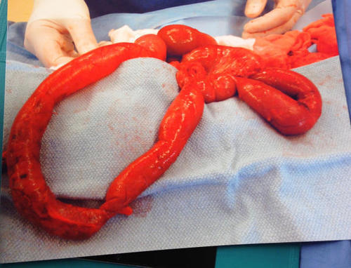
Rapidly place a nasogastric tube to try to decompress the stomach. It doesn’t always work because the angles at the hiatus are not what they usually are. But if it does, the patient will feel better immediately. You may be able to avoid intubating them… for a few minutes. This is a surgical problem, and commonly involves injury to other abdominal structures, especially the spleen. Order up some blood, and rapidly complete your evaluation. A pelvis x-ray is in order, because fractures are common after full or partial ejection through the window. No other imaging is necessary. The send your patient off to the OR for a thorough exploration and repair. Now you can intubate.

I’ve seen this injury three times after partial ejection, and always involving the driver (left side, makes sense). Any time you have left sided abnormalities after blunt trauma that don’t respond to a chest tube, think about this problem.

What The Heck? The original presentation

What The Heck? Part 2

Thanks to all who tweeted, commented and emailed their suggestions! The case involves a partially ejected patient who is brought to the ED with respiratory distress and diminished lung sounds on the left. 

Sounds easy, right? But remember, this is the Trauma Professional’s Blog! I want you to be prepared for things that are a little outside the ordinary. No zebras here, but stuff you could actually see.

First, every trauma activation patient gets supplemental oxygen as the festivities continue. You need to quickly figure out if this is an airway, breathing, or circulation problem. Yes, circulation. Major torso vascular injuries and tamponade can cause respiratory distress. However, we would not expect the blood pressure to be anywhere near normal.

So check the airway to make sure there is no foreign material there. Check the trachea for position. This is one of those classic test results in medicine: if the trachea is deviated, they most likely have a tension pneumothorax. But if it’s not, that doesn’t necessarily mean they don’t. In this case, the trachea is in its usual place, but don’t count tension physiology out yet.

Double check the breath sounds. You confirm that they are nearly absent on the left. What to do next?

You must presume some major problem on the left: large hemo- or pneumothorax, or a tension pneumothorax. Since your patient is physiologically abnormal, you cannot wait to get a chest x-ray. You have to deal with the breathing problem right away. The correct answer is to needle the left chest, then follow immediately with a chest tube.

You do so, and both procedures go smoothly. The chest tube fogs with exhalation, and there is a small amount of blood (100cc) that drains into the collection system. But your patient does not look or feel any better! Oxygen saturations are still in the low 80′s, and he remains dyspneic. As you were finishing the chest tube, the radiology tech snapped a quick chest x-ray, and the result will be up in two minutes.

Now what? Your choices are:

  • Intubate
  • Insert another chest tube
  • Package the patient and run to the OR
  • Wait for the chest x-ray

Again, tweet, comment or email. What is wrong, and why didn’t the chest tube work? What is the ideal next move? Answer tomorrow.

What The Heck? The original presentation

What The Heck? Pediatric Abdomen: The Answer

By now, you know the story. Sick little girl who comes to your ED with a rigid abdomen. She’s been previously healthy, and there is no history of trauma.

Here’s the surgical specimen again:

This is a loop of small bowel. It it very inflamed, and you can see a darker color on the portion that is at the left of the picture. This is an area of necrosis, indicating that this portion lost its blood supply some time ago, possibly even a day or two.

A number of readers guessed volvulus or internal hernia, which are on the usual list of differential diagnoses.

But hey, this is the Trauma Professional’s Blog!

Look again carefully at that loop of bowel on the left. There is no mesentery attached to it! This is a classic buckethandle injury. And the only way to get it is from blunt force, typically a car crash. There were also low grade lacerations of the liver and spleen.

What’s the diagnosis now? Non-accidental trauma!

On closer questioning, the father’s story began to change when confronted with this information. As the child recovered from surgery, she underwent a workup and was found to have a number of healed and healing rib fractures of various ages. Child protective services was involved, and the father ultimately admitted to repeatedly striking her in the abdomen in a fit of rage.

Bottom line: Healthy children who are abruptly found to be very ill (or in cardiac arrest) have a high likelihood of non-accidental trauma in addition to the usual medical and surgical culprits. Never lose sight of that, and always maintain some suspicion, no matter how nice the parents seem to be. Treat the child, but always be cognizant of their social/domestic situation!

What The Heck? Pediatric Abdomen Part 2

Yesterday, a child was brought to your ED who looked bad and felt bad, to the point you are convinced she has peritonitis. If the abdominal exam is convincing enough, there is no need for diagnostic imaging It would only serve to add time and radiation to the equation. 

If you had really insisted on getting something like a CT, I would have told you that it just showed nonspecific distension and a small amount of free fluid. Helpful, right? And that at the age of 34 she would develop lymphoma for no apparent reason.

Your surgery team takes this patient immediately to the OR and finds this:

So now my questions are:

  • What is going on here? What is this?
  • What is really going on here? What’s the problem?

Please Tweet and leave emails or comments with your guesses. Discussion and answers tomorrow!

What The Heck? Pediatric Abdomen

Here’s an interesting case for you to think about. A three year old female is brought to your ED with a three day history of abdominal pain which has been getting worse. She started vomiting yesterday, and hasn’t wanted to eat for two days. The child has been completely healthy until now. The parents deny any history of trauma.

On exam, the child appears to be ill. She has a distended abdomen bordering on rigidity, and is markedly tender. A FAST exam is performed, which shows a small amount of fluid only in the pelvis.

Here are my questions for you:

  • What tests would you like to order?
  • What is going on here?

Remember, this is the Trauma Professional’s Blog, after all!