Tag Archives: VTE

Enoxaparin And anti-Xa Levels: Who Cares? Part 2

In my last post, I reviewed a study that looked at monitoring factor anti-Xa for the purpose of just “hitting the number.” Not very convincing. Today, I’ll review one that studied a reasonable outcome, the actual occurrence of VTE in patients.

This was another small, prospective study at a busy Level I trauma center. The outcomes that were analyzed included LOS, transfusion requirement, hematocrit on discharge, and diagnosis of deep venous thrombosis (DVT) or pulmonary embolism (PE). Only the last two of these make sense, especially for this small study. (205 patients in two 10 month periods).

At this center, all trauma patients are started on enoxaparin, regardless of injury severity. And all patients have sequential compression devices applied unless contraindicated by their injuries. Patients were included if the were administered 3 consecutive enoxaparin doses and had a trough anti-Xa level measured an hour before the fourth dose. If the trough was less than 0.1 IU/ml, dosing was adjusted until it rose to > 0.2 IU/ml. Outcomes were compared to historical controls from the prior year.

Here are the factoids:

  • A total of 87 study patients were enrolled in 10 months.  However, this represents only about 15% of trauma admissions to the center. Why were so few eligible for inclusion?
  • 84% of study patients did not “hit the number” with 30mg bid dosing (again!)
  • They were compared to 118 control patients who received enoxaparin during the same 10 month period, a year earlier
  • Screening by duplex ultrasound was only done for “clinical suspicion” of DVT or PE. No routine screening. And we know how reliable clinical suspicion can be.
  • 84% of patients were not at their anti-Xa goal when the first trough was done. Most of these patients needed 40mg bid to “hit the number.”
  • DVT and PE occurrences were “significantly lower” in the dose adjusted group compared to historical controls (1.1% vs 7.6%). Now this is a difference between only 1 adjusted patient and 9 controls, and the p value barely made it at 0.046.
  • Proximal DVT occurred  in no adjusted patients vs 2 controls (not significant)
  • PE occurred in no adjusted patients and 1 control (not significant)
  • Distal DVT occurred in 1 adjusted patient and 6 controls (not significant

Bottom line: This is yet another (very) small study. It also demonstrates why you must read the study, not just the abstract! The study group was a fraction of all of the patient admitted, even though all patients supposedly received prophylaxis. The attending physicians decided when to start dosing, and this varied from 0 to 4 days. Screening was ordered only if there was some kind of clinical suspicion for DVT or PE, and the details were not spelled out. 

For all these reasons, there are many, many opportunities for bias. But probably the most important problem is the statistics. I always worry when the p value for a numerical difference barely reaches 0.05, especially when the actual numbers look to be far apart. It is usually an indicator of small study size.

But in this case, the breakdown of VTE location is critical. The sums of the distal, proximal, and pulmonary occurrences show a p value difference just under 0.05. But when you compare study vs control for each, the bulk of the numbers are due to distal DVT.  The literature does not convincingly support prophylaxis for distal DVT, and we do not even treat it at my center. We continue surveillance to make sure it doesn’t creep up into the popliteal arteries.

This is yet another weak study trying to make the case for anti-Xa monitoring that doesn’t pass muster. Again, we see that 30mg bid doesn’t “hit the number” without adjustment. But we also haven’t shown that hitting that magic number of 0.2 IU/ml (peak or trough) by adjusting the dose makes a difference either.

But we continue to try. In my next post, we’ll look at another recently published study on the same topic.

Related posts:

Reference: Association between enoxaparin dosage adjusted by anti-factor Xa trough level and clinically evident venous thromboembolism after trauma. Jama Surg. Published online ahead of print July 6, 2016.

Enoxaparin And anti-Xa Levels: Who Cares? Part 1

Several papers have been published in recent years analyzing the process of fine-tuning venous thromboembolism (VTE) prophylaxis with enoxaparin. My own hospital has (or had) a protocol in place to automatically draw anti-Xa levels after the third enoxaparin dose in select patients. What is the science behind this concept? It looks like that’s a popular question these days.

Enoxaparin interacts with antithrombin III, turning off a number of factors further down in the clotting cascade. As part of the process, it inactivates Factor Xa, which is easily measurable by a simple blood test. This is very helpful, since PT and PTT are not affected by enoxaparin.

The paper I will discuss today postulated that many patients are “sub-therapeutic” given the usual dosing regimen of 30mg bid. They primarily focused on “hitting the number”, meaning achieving an anti-Xa level > 0.2 IU/ml.

Patients at a single Level I trauma center were enrolled, receiving standard dose enoxaparin and undergoing duplex screening within 48 hours of admission, and again during the first week in hospital. Anti-Xa levels were drawn four hours after the third dose (peak level) and one hour before the fourth dose (trough level).

Here are the factoids:

  • Of 164 patients enrolled, only 61 patients remained in the study. A total of 103 (63%) were excluded because blood draws or screening studies were not done correctly. (!!)
  • 70% of patients had sub-therapeutic enoxaparin dosing based on anti-Xa peak levels
  • The subtherapeutic patients tended to be males, with “higher body weight.” The reality was that the therapeutic patients weighed 71kg and the non-therapeutic men 88kg. But BMI was only 25 and 29, respectively, and was not significantly different.
  • There were 3 VTEs detected during the study, all receiving the initial 30mg dose of enoxaparin. Two of the three had therapeutic anti-Xa levels.
  • No bleeding complications were observed in patients who had their enoxaparin dose adjusted upward

Bottom line: It’s really hard to take anything away from this study at all! Well, we can certainly see that the research group had a tough time adhering to their own protocol, losing two thirds of their study group. This throws the accuracy of the data on the remaining subjects into doubt given the very low numbers.

It would appear that many patients did not achieve their magic number of 0.2 IU/ml for anti-Xa when receiving the standard enoxaparin dose. So what? VTE occurred essentially equally in both groups, but really can’t be interpreted either due to the low numbers.

So basically, this paper is just telling us how many of their patients don’t hit the magic number. Not if that number has any implications on real outcomes, like DVT, PE, or mortality. But if you only read the title or abstract, you might think so!

Tomorrow, I’ll review a paper on anti-Xa that takes a different approach. Just about as successfully.

Related posts:

Reference: Dose adjusting enoxaparin is necessary to achieve adequate venous thromboembolism prophylaxis in trauma patients. J Trauma 745(1):128-135, 2013.

Predicting VTE Risk In Children

There’s a lot of debate about if and at what age injured children develop significant risk for venous thromboembolism (VTE). In the adult world, it’s a little more clear cut, and nearly every patient gets some type of prophylactic device or drug. Kids, we’re not so certain about at all.

The Children’s Hospital of Wisconsin tried to tease out these factors to develop and implement a practice guideline for pediatric VTE prophylaxis. They prospectively reviewed over 4000 pediatric patients admitted over a 6 year period.

It looks like the guideline was developed using some or all of this data, then tested using regression models to determine which factors were significant. The guideline was then tweaked and a final model implemented.

Here are the factoids:

  • 588 of the patients (14%) were admitted to the ICU, and 199 of these were identified as high risk by the guidelines
  • Median age was 10 (this is always important in these studies)
  • VTE occurred in 4% of the ICU patients, and 10% of the high risk ones
  • Significant risk factors included presence of central venous catheter, use of inotropes, immobilization, and GCS < 9

Bottom line: This abstract confuses me. How were the guidelines developed? What were they, exactly? And the results seem to pertain to the ICU patients only. What about the non-ICU kids? The abstract just can’t convey enough information to do the study justice. Hopefully, the oral presentation will explain all.

I prefer a very nice analysis done at the Oregon Health Science University in Portland. I wrote about this study earlier this year. The authors developed a very useful calculator that includes most of the risk factors in this model, and a few more. Input the specific risks, and out comes a nice score. The only issue is, what is the score threshold to begin prophylaxis and monitoring? Much more practical (and understandable) than this abstract. Check it out at the link below.

Related post:

References:

  1. Evaluation of guidelines for injured children at high risk for VTE: a prospective observational study. AAST 2016, Paper 68.
  2. A Clinical Tool for the Prediction of Venous Thromboembolism in Pediatric Trauma Patients. JAMA Surg 151(1):50-57, 2016.

Battle of the Heparins: Unfractionated vs Low Molecular Weight

Most trauma programs tend toward using low molecular weight heparin (LMWH) products for VTE prophylaxis over plain, old-fashioned unfractionated heparin (UH). How did this happen? LMWH is more expensive than UH, and there is precious little high quality research supporting it.

But, LMWH is very convenient, as it only needs to be given only once or twice daily via subq injection, whereas UH is given as a continuous infusion or subq three times a day. And a fair amount of lower quality data suggests that it is effective in decreasing deep venous thrombosis (DVT) and pulmonary embolism (PE).

This abstract comes from Sunnybrook in Toronto. The authors used sophisticated statistical models to compare centers that predominantly use LMWH to prevent VTE vs those that use UH.

Here are the factoids:

  • This was a huge data analysis from the ACS Trauma Quality Improvement Program database (~ 110,000 records from 214 trauma centers)
  • LMWH was most commonly used, 74% of the time
  • Patients who were more likely to need rapid reversal were more often given UH (older patients, severe TBI, early intracranial interventions)
  • Pulmonary embolism was significantly lower with LMWH (1.8% vs 2.4%)
  • This significant effect was present across all subgroups, including patients with shock, blunt multisystem injury, penetrating trunk injury, isolated orthopedic injury, and severe TBI
  • Trauma centers that predominantly used LMWH had significantly lower PE rates compared to UH (1.2% vs 1.8%)

Bottom line: Even given the vagaries of using huge, retrospective database reviews, this is pretty good data. The use of LMWH appears to be superior to UH in reducing the incidence of pulmonary embolism. It does not prevent it completely. But it’s a good start.

What the authors do not say, and I am curious about, is the impact on DVT. That is a much more common problem than PE. Was there any difference? Did they run out of room to comment on it in the abstract? I kind of doubt it. The devil will be in the details. Listen in on the presentation at the meeting!

Reference: Efficacy of low molecular weight heparin vs unfractionated heparin to prevent pulmonary embolism following major trauma: results from the American College of Surgeons Trauma Quality Improvement Program. AAST 2016 Paper #5.

How To Predict Venous Thromboembolism In Pediatric Trauma

As with adults a decade ago, the incidence of venous thromboembolism (VTE) in children is now on the rise. Whereas adult VTE occurs in more than 20% of adult trauma patients without appropriate prophylaxis, it is only about 1% in kids, but increasing. There was a big push in the early 2000′s to develop screening criteria and appropriate methods to prevent VTE. But since the incidence in children was so low, there was no impetus to do the same for children.

The group at OHSU in Portland worked with a number of other US trauma centers, and created some logistic regression equations based on a large dataset from the NTDB. The authors developed and tested 5 different models, each more complex than the last. They ultimately selected a model that provided the best fit with the fewest number of variables.

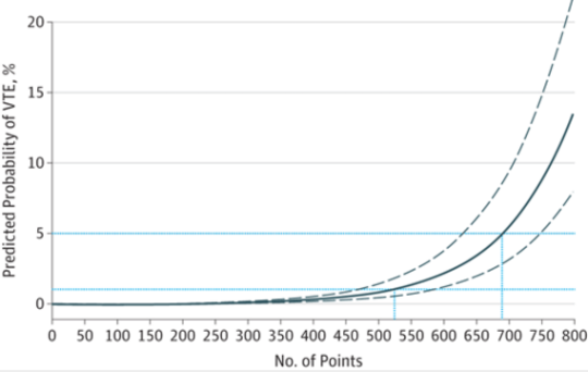
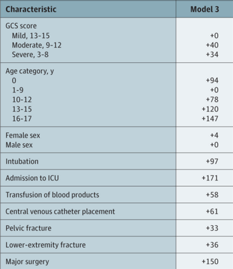
The tool consists of a list of risk factors, each with an assigned point value. The total point value is then identified on a chart of the regression equation, which shows the risk of VTE in percent.

Here are the factors:

Note that the highest risk factors are age >= 13, ICU admission, and major surgery.

And here is the regression chart:

Bottom line: This is a nice tool, and it’s time for some clinical validation. So now all we have to do is figure out how much risk is too much, and determine which prophylactic tools to use at what level. The key to making this clinically usable is to have a readily available “VTE Risk Calculator” available at your fingertips to do the grunt work. Hmm, maybe I’ll chat with the authors and help develop one!

Reference: A Clinical Tool for the Prediction of Venous Thromboembolism in Pediatric Trauma Patients. JAMA Surg 151(1):50-57, 2016.