Tag Archives: TBI

How Long Should We Watch Intracerebral Hemorrhage?

Patients with traumatic brain injury (TBI) severe enough to cause bleeding are usually admitted to the hospital for observation and in many cases, repeat CT scanning. Those with small intracranial hemorrhages (ICH) may experience progression of the bleeding, and a small percentage of cases may need operative intervention (1-3%). Questions we typically face are, how long should we watch for progression, and how often should we scan?

A retrospective cohort study was carried out at UMD-NJ, looking for answers for a specific subset of these patients. Specifically, they had to have a mild blunt TBI (loss of consciousness and/or retrograde amnesia, GCS in the ED of 13-15) and a positive head CT. They classified any type of hemorrhage into or around the brain as positive. 

During a 3 year period, 474 adults were enrolled but only 341 were eligible for the study. They were excluded due to previous injury, presence of a mass (not trauma), need for immediate neurosurgical intervention, or failure to get a second CT scan. The authors found:

  • 7% of patients were taking anticoagulants! This is surprisingly high. Interestingly, 15 were subtherapeutic, 3 were therapeutic and 2 were supratherapeutic.
  • Subarachnoid hemorrhage was the most common finding on CT (54%). Intraparenchymal hemorrhage was next most common (48%) Many patients had more than one type of bleed.
  • The injury worsened between the first and second scans in 31% of patients. This number increased to 46% in patients taking anticoagulants. 
  • About 97% of bleeds stopped progressing by 24 hrs post-injury.

Bottom line: Most centers are probably overdoing the observation and repeat scan thing. More than two thirds of bleeds are stable by the first scan (first and second scans identical), and nearly all stop progressing within 24 hours. It’s very likely that patients who are not on anticoagulants and who have a stable neuro exam and stable symptoms can get just one scan and 24 hours of observation. Persistent headache, nausea, failure to ambulate well, or other symptoms warrant a repeat scan and longer observation.

Related posts:

Reference: The temporal course of intracranial haemorrhage progression: How long is observation necessary? Injury 43(12):2122-2125, 2012.

Can TBI Be Managed Without Neurosurgical Consultation?

The standard of care in most high level trauma centers is to involve neurosurgeons in the care of patients with significant traumatic brain injury (TBI). However, not all hospitals that take care of trauma patients have immediate availability of this resource. A paper to be presented at the upcoming EAST meeting looked at management of these patients by acute care surgeons.

The authors retrospectively reviewed all patients who had a TBI and positive head CT managed with or without neurosurgery consultation over a two year period. Although the authors were from the University of Arizona, a Level I ACS trauma center, the abstract does not explicitly state whether the patients were seen in their hospital or another lower level one.They matched the patients with and without neurosurgical consultation for age, GCS, AIS-Head and presence of skull fracture and intracranial hemorrhage.

A total of 90 patients with and 90 patients without neursurgical involvement were reviewed. Here are the interesting findings:

  • Hospital admission rate was identical for both groups (87-90%)
  • ICU admission was significantly higher if neurosurgeons were involved (20% vs 41%)
  • Repeat head CT was ordered more than 3 times as often by neurosurgeons (20% vs 72%)
  • Post-discharge head CT was ordered more often by neurosurgeons, but was not significantly higher (5% vs 12%)

Nothing is said about complications or mortality, or whether neurosurgeons were available in case things went awry.

Bottom line: This abstract raises an interesting question: can surgeons safely manage select patients with intracranial injury? The answer is probably yes, although this abstract is not complete enough to fully support the idea. The majority of patients with mild to moderate TBI with small intracranial bleeds do well despite everything we throw at them. And it appears that surgeons use fewer resources managing them than neurosurgeons do. The keys to being able to use this type of system are to identify at-risk patients who really do need a neurosurgeon early, and having a quick way to get the neurosurgeon involved (by consultation or hospital transfer). As neurosurgery involvement in acute trauma declines, this concept will become more and more pertinent.

Related posts:

Reference: The acute care surgery model: managing traumatic brain injury without an inpatient neurosurgical consultation. EAST Annual Scientific Assembly, Paper 10, January 2013.

The Value Of Repeated Head CT

Repeating the head CT in patients with head injury has almost become routine. This practice varies greatly and depends on the neurosurgeon’s preference in many centers. It occurs most commonly when there is blood inside the skull, any type of blood. But does this practice make sense? Sure, if you’ve got a small epidural it seems reasonable. But what about that wisp of subarachnoid blood?

Another paper being presented at the Congress of Neurologic Surgeons this fall describes a review of 445 cases at their hospital and a meta-analysis of 15 studies in the literature. The authors looked at the practice of repeat CT scanning with respect to the good old clinical exam. They stratified all patients who underwent an intervention after repeat CT into two groups, based on changing clinical exam or CT findings.

They found that a significant number of patients required some management change based on deteriorating neurologic exam, whereas very few required it based on the repeat CT. The authors concluded that it is not necessary to rescan a head trauma patient if their neurologic status is stable or improving. 

Bottom line: This is preliminary data, and it is only available in abstract form, so don’t change your practice yet. We need more information on how many patients were reviewed and how good the meta-analysis was. However, you should begin to question whether rescanning everyone is necessary or prudent. Not all head injuries are alike, and some of the trivial ones, like subarachnoid blood in a young adult probably don’t need a repeat scan. More to come when this is presented and/or published.

Reference: The Value of Scheduled Repeat Cranial Computed Tomography Following Mild Head Injury: Single Center Experience and Meta-Analysis. Paper 152, presented at the Congress of Neurologic Surgeons, October 2012.

Subdural Hematomas and Hygromas Simplified

There’s a lot of confusion about subdural pathology after head trauma. All subdural collections are located under the dura, on the surface of the brain. In some way they involve or can involve the bridging veins, which are somewhat fragile and get more so with age.

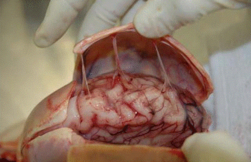
Head trauma causes a subdural hematoma by tearing some of these bridging veins. Notice how thick the dura is and how delicate the bridging veins are in the image below.

When these veins tear, bleeding ensues which layers out over the surface of the brain in that area. If the bleeding does not stop, pressure builds and begins compressing and shifting the brain. A subdural hematoma is considered acute from time of injury until about 3 days later. During this time, it appears more dense than brain tissue.

After about 3-7 days, the clot begins to liquefy and becomes less dense on CT. Many hematomas are reabsorbed, but occasionally there is repeated bleeding from the bridging veins, or the hematoma draws fluid into itself due to the concentration gradient. It can enlarge and begin to cause new symptoms. During this period it is considered subacute.

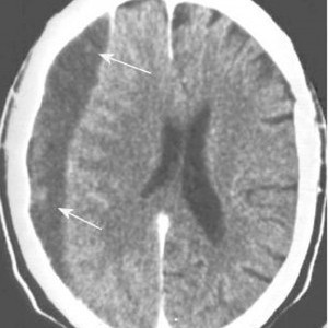
It moves on to a more chronic stage over the ensuing weeks. The blood cells in it break down completely, and the fluid that is left is generally less dense than the brain underneath it. The image below shows a chronic subdural (arrows).

Hygromas are different, in that they are a collection of CSF and not blood. They are caused by a tear in the meninges and allow CSF to accumulate in the subdural space. This can be caused by head trauma as well, and is generally very slow to form. They can lead to slow neurologic deterioration, and are often found on head CT in patients with a history of falls, sometimes in the distant past. CT appearance is similar to a chronic subdural, but the density is the same as CSF, so it should have the same appearance as the fluid in the ventricle on CT.

Related posts:

Return To Baseline After Concussion

Here’s another interesting paper that was presented at the Congress of Neurological Surgeons. There’s a lot of attention being focused on the incidence and management of concussion during sporting events. An international Concussion in Sport Group has been meeting for over 10 years, contemplating classification and management of this injury. They are considering using age to modify management of concussion in young athletes.

The authors looked at their own experience with 200 adolescent and young athletes. They stratified by age (younger = 13-16 year olds, older = 18-22 year olds), with 100 in each group. They matched them by number of previous concussions, and all underwent baseline and post-concussion ImPACT testing. They specifically looked at the number of days needed to return to baseline.

Interestingly, they identified significant differences in recovery time. And strangely enough, the older players did better than the younger ones. Overall, 90% returned to baseline within a month. But the younger players took 2-3 days longer to recover than the older ones. 

Bottom line: Looks like the Concussion in Sport Group is right on! Usually in trauma, older folks do worse than younger ones, so we tend to treat them more carefully. Not so in youngsters with concussions. Sports medicine physicians need to realize that the younger brain takes longer to recover, and they should err on the safe side and keep them out of the game longer. Objective testing to help predict return to play is extremely helpful.

Related post:

Reference: Sport-Related Concussion and Age: Number of Days to Neurocognitive Baseline. Oral presentation 145 – Congress of Neurological Surgeons 2012.