Tag Archives: pulmonary edema

Flash Pulmonary Edema After Chest Tube Insertion

You are seeing a young man in the emergency department who gives a history of falling two days ago. He experienced chest pain at the time which has persisted, but he did not immediately seek medical care. He has noticed that he now gets winded when walking quickly or climbing stairs, and describes pleuritic chest pain.

He presents to your emergency room and on exam has a bruise over his left lateral chest wall. Subcutaneous emphysema is present, and breath sounds are absent. Chest x-ray shows a complete pneumothorax on the left.

You carefully prepare and insert a chest tube in the usual position. A significant rush of air occurs, which tapers off over 15 seconds. Here is the followup image:
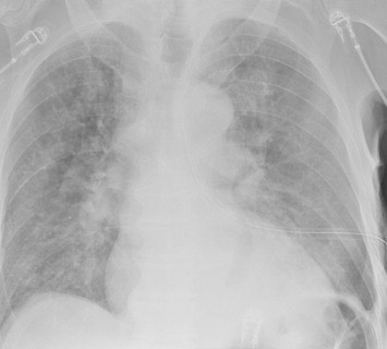
About 10 minutes later you are called to his room because he is complaining of dyspnea and his oxygen saturation has decreased to 86%. Breath sounds are somewhat decreased and the tube appears to be functioning properly. You immediately obtain another chest x-ray:

What just happened? This is a classic case of unilateral “flash” pulmonary edema after draining the chest cavity. This phenomenon was first described in 1853 in a patient who had just undergone thoracentesis. It is very uncommon, but seems to occur after rapid drainage of air or fluid from the chest cavity.

Here are some interesting factoids from case reports:

  • It occurs more often in young men
  • It is most common when draining large hemo- or pneumothoraces
  • Rapid drainage seems to increase the incidence
  • It is likely due to increased pulmonary capillary permeability from inflammatory mediators or changes in surfactant
  • Symptoms typically develop within an hour after drainage

What should you do? First, if you are draining a large collection of air or blood, do it slowly. Clamp the back end of the chest tube prior to insertion (you should always do this if you value your shoes) and use it to meter the amount of fluid or air released. I typically let out about 300cc of fluid, then wait a minute and repeat until all the blood has been drained. For air, vent it for 10 seconds, then wait a minute and repeat.

In patients at high risk for this condition, apply pulse oximetry and follow for about an hour. If they still look and feel great, nothing more need be done.

References:

  • Fulminant Unilateral Pulmonary Edema After Insertion of a Chest Tube. Dtsch Arztebl Int 105(50):878-881, 2008.
  • Reexpansion pulmonary edema after chest drainage for pneumothorax: A case report and literature overview. Respir Med Case Rep 14:10-12, 2015.
  • Re-expansion pulmonary edema following thoracentesis, Can Med Assn J 182(18):2000-2002, 2010.
Pulmonary Edema After Chest Tube Insertion

Re-expansion pulmonary edema is an uncommon event after chest tube insertion. Typically, patients have had symptoms of pneumothorax for several days, usually 3 or more. It occurs most often if a large amount of air (or blood) is evacuated at once. The patient will typically become symptomatic within an hour, with decreased oxygen saturation and subjective breathing difficulty.

Although the mechanism is not entirely clear, it appears that the small blood vessels in the lung become more permeable if they are collapsed for an extended period. Mechanical stress from rapid re-expansion further damages the vessels, allowing them to leak. This leads to oxygenation and ventilation problems if severe. 

Practical tips:

  • Check the history. Most of these patients have had their pneumothorax for 3 or more days.
  • Check the xray. Complete pneumothorax (or large hemothorax) puts the patient at high risk.
  • Modify your chest tube insertion technique. Clamp the distal end of the tube so the pneumothorax is not evacuated suddenly as the tube goes in.
  • Modify the collection system. Do not use suction initially; only set up for water seal. Clamp the tubing on the patient side. Every 10-15 minutes release the clamp and briefly let some of the air out of the chest, then reclamp. Repeat this until all air has bubbled through the water seal chamber. 
  • Watch your patient. If they cough excessively, start to desaturate or become dyspneic, get your respiratory adjuncts. Give higher inspired oxygen by appropriate means, and consider BiPap or CPAP. In extreme cases intubation may be needed. If the patient does not have any difficulties after about an hour, connect the collection system to suction and proceed as you normally would.

Reference: Reexpansion pulmonary edema. Ann Thoracic Cardiovasc Surg 14:205-209, 2008.