Tag Archives: Penetrating trauma

Managing Penetrating Injuries: Some Practical Tips

Although penetrating injuries are a relatively uncommon mechanism at most trauma centers, they are more likely than not to injure deeper structures. Key decisions need to be made quickly during the initial evaluation in order to provide the best care.

Here are some practical tips:

  • Penetrating injuries to just about anything but the extremities should activate your trauma team.
  • If your patient is hypotensive, they will need to go to the OR. You can certainly start infusing some fluid or blood, but a lot leaked out before they got to you, indicating that the leak needs to be surgically fixed. No exceptions.
  • All hypotensive patients require activation of your massive transfusion protocol and consideration of giving tranexamic acid (TXA).
  • If your patient is normotensive, you have the luxury of evaluating them more thoroughly. But don’t lose your sense of urgency. Assume they are dying until you prove otherwise.
  • Complete your secondary survey. Don’t skimp on the exam and always look at the back. If your patient ends up on an OR table, it may be the only time you get to look at it for quite some time.
  • Get a single x-ray of the affected area, even if you need to go to the OR quickly. This can help plan your operation, and may drive you to explore areas you had not considered.
  • Before shooting the x-ray, mark any and all entry and exit points. This will help to predict the trajectory and any injured structures.
  • Use small markers, but not too small. Most radiology departments have small arrows, which are ideal. Dots are too small and may not show up well on plain images. But be aware that some markers may be too dense for CT, causing artifacts that may obscure pathology.
  • Watch out for your own safety! Somebody was trying to kill your patient, and they may show up at your hospital to try to finish the job. Make sure your ED and inpatient areas take appropriate security precautions.

How To Read A Stab Wound

Most emergency departments do not see much penetrating trauma. But it is helpful to be able to learn as much as possible from the appearance of these piercing injuries when you do see them. This post will describe the basics of reading stab wounds.

Important: This information will allow some basic interpretation of wounds. It will not qualify you as a forensics expert by any means. I do not recommend that you document any of this information in the medical record unless you have specific forensic training. You should only write things like “a wound was noted in the midepigastrium that is 2 cm in length.” Your note can and will be used in a court of law, and if you are wrong there can be significant consequences for the plaintiff or the defendant. This information is for your edification only.

1. What is the length of the wound? This does not necessarily correspond to the width of the blade. Skin stretches as it is cut, so the wound will usually retract to a length that is shorter than the full width of the blade.

2. Is the item sharp on one side or both? This can usually be determined by the appearance of the wound. A linear wound with two sharp ends is generally a two sided knife. A wound with one flat end and one sharp end is usually from a one-sided weapon. The picture below shows a knife wound with one sharp side.

3. Is there a hilt mark? This can usually be detected by looking for bruising around the wound. The picture below shows a knife wound with a hilt mark.

4. What is the angle? If both edges are symmetric, the knife went straight in. If one surface has a tangential appearance, then the knife was angled toward that side. You can approximate the direction of entry by looking at the tangential surface of the wound edge. In this example, the blade is angling upward toward the right.

5. How deep did it go? You have no way of knowing unless you have the blood stained blade in your possession. And yes, it is possible for the wound to go deeper than the length of the knife, since the abdominal wall or other soft tissues can be pushed inwards during the stab.

How Good Is The Spine Exam In Penetrating Injury?

Examination of the spine in trauma patients is typically not very helpful. We always look for stepoffs. swelling and tenderness, but the correlation with actual injury is poor. A recent paper presented at the American Medical Student Association Annual Convention showed that it actually can be helpful in victims of penetrating injury.

A prospective study of 282 patients was carried out at a Level I Trauma Center, specifically focusing on penetrating trauma. Half had gunshot wounds, and 8% sustained spinal injury with one third left with permanent disability. Stab wounds never led to a spinal cord injury. The most common patterns for cord injury in gunshot wounds was a single shot to the head or neck, or multiple shots to the torso.

The examiners looked for pain, tenderness, deformity and neurologic deficit. They found that the sensitivity was 67%, the specificity was 90%, the positive predictive value was 95% and the negative predictive value was 46%. These numbers are much better than those found during spine examination after blunt trauma. They also determined that prehospital immobilization after penetrating injury would not have helped, which I have also written about here.

Bottom line: A good spine exam in victims of penetrating trauma can accelerate definitive management prior to defining the exact details of the injury with radiographic or MRI imaging. This is particularly helpful in patients who present to non-trauma centers, where imaging or image interpretation may not be readily available.

Reference: American Medical Student Association (AMSA) 60th Annual Convention: Abstract 26: Presented March 11, 2010

Off-Label Use of the Foley (Urinary) Catheter

Foley catheters are a mainstay of medical care in patients who need control or measurement of urine output. Leave it to trauma surgeons to find warped, new ways to use them!

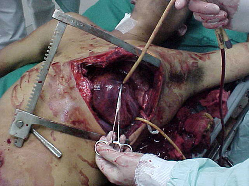
Use of these catheters to tamponade penetrating cardiac injuries has been recognized for decades (see picture, 2 holes!). Less well appreciated is their use to stop bleeding from other penetrating wounds.

Foley catheters can be inserted into just about any small penetrating wound with bleeding that does not respond to direct pressure. (Remember, direct pressure is applied by one or two fingers only, with no flat dressings underneath to diffuse the pressure). Arterial bleeding, venous bleeding or both can be controlled with this technique. 

In general, the largest catheter with the largest possible balloon should be selected. It is then inserted directly into the wound until the entire balloon is inside the body. Inflate the balloon using saline until firm resistance is encounted, and the bleeding hopefully stops. Important: be sure to clamp the end of the catheter so the bleeding doesn’t find the easy way out!

Use of catheter tamponade buys some time, but these patients need to be in the OR. In general, once other life threatening issues are dealt with in the resuscitation room, the patient should be moved directly to the operating room. In rare cases, an angiogram may be needed to help determine the type of repair. However, in the vast majority of cases, the surgeon will know exactly where the injury is and further study is not needed. The catheter is then prepped along with most of the patient so that the operative repair can be completed.

Related posts:

Best Of: Off-Label Use of the Foley Catheter

Foley catheters are a mainstay of medical care in patients who need control or measurement of urine output. Leave it to trauma surgeons to find warped, new ways to use them!

Use of these catheters to tamponade penetrating cardiac injuries has been recognized for decades (see picture, 2 holes!). Less well appreciated is their use to stop bleeding from other penetrating wounds.

Foley catheter in heart

Foley catheters can be inserted into just about any small penetrating wound with bleeding that does not respond to direct pressure. (Remember, direct pressure is applied by one or two fingers only, with no flat dressings underneath to diffuse the pressure). Arterial bleeding, venous bleeding or both can be controlled with this technique. 

In general, the largest catheter with the largest possible balloon should be selected. It is then inserted directly into the wound until the entire balloon is inside the body. Inflate the balloon using saline until firm resistance is encounted, and the bleeding hopefully stops. Important: be sure to clamp the end of the catheter so the bleeding doesn’t find the easy way out!

Use of catheter tamponade buys some time, but these patients need to be in the OR. In general, once other life threatening issues are dealt with in the resuscitation room, the patient should be moved directly to the operating room. In rare cases, an angiogram may be needed to help determine the type of repair. However, in the vast majority of cases, the surgeon will know exactly where the injury is and further study is not needed. The catheter is then prepped along with most of the patient so that the operative repair can be completed.

Related posts: