Tag Archives: IV

IV Contrast In Trauma Imaging

We use CT scanning in trauma care so much that we tend to take it (and its safety) for granted. I’ve written quite a bit about thoughtful use of radiographic studies to achieve a reasonable patient exposure to xrays. But another thing to think about is the use of IV contrast.

IV contrast is a hyperosmolar solution that contains some substance (usually an iodine compound) that is radiopaque to some degree. It has been shown to have a significant impact on short-term kidney function and in some cases can cause renal failure.

Here are some facts you need to know:

  • Contrast nephrotoxicity is defined as a 25% increase in serum creatinine, usually within the first 3 days after administration
  • There is usually normal urine output and minimal to no proteinuria
  • In most cases, renal function returns to normal after 3-4 days
  • Nephrotoxicity almost never occurs in people with normal baseline kidney function
  • Large or repeated doses given within 72 hours greatly increase risk for toxicity
  • Old age and pre-existing diabetic renal impairment also greatly increase risk

If you must give contrast to a patient who is at risk, make sure they are volume expanded (tough in trauma patients), or consider giving acetylcysteine or using isosmolar contrast (controversial, may still cause toxicity).

Bottom line: If you are considering contrast CT, try to get a history to see if the patient is at risk for nephrotoxicity. Also consider all of the studies that will be needed and try to consolidate your contrast dosing. For example, you can get CT chest/abdomen/pelvis and CT angio of the neck with one contrast bolus. Consider low dose contrast injection if the patient needs formal angiographic studies in the IR suite. Always think about the global needs of your patient and plan accordingly (and safely).

Related posts:

Reference: Contrast media and the kidney. British J Radiol 76:513-518, 2003.

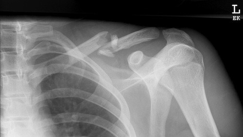
What Do You Think? Insert A Subclavian IV Near A Clavicle Fracture?

Someone asked me an interesting question: If I have a patient with a clavicle fracture, is it okay to insert a subclavian line on that side?

Don’t try to look it up. As far as I’ve been able to determine, there is no literature on this simple little question. For most people, the first reaction is “No way!” But think about it a bit, and see if you can come up with any reasons it might be preferred.

Please post your comments, or tweet them out. I’ll answer this pesky one tomorrow.

The EMS Second IV In Trauma

One of the critical maneuvers that EMS providers perform is establishing initial vascular access. This IV is important for administering medications and for initiating volume resuscitation in trauma patients. Prehospital Trauma Life Support guidelines state that every trauma patient should receive two large bore IV lines. But is this really necessary?

The upside of having two IVs in the field is that the EMS provider can give lots of volume. However, a growing body of literature tells us that pushing systolic blood pressure up to “normal” levels in people (or animals) with an uncontrolled source of bleeding can increase mortality and hasten coagulopathy.

The downside of placing two lines is that it is challenging in a moving rig, sterility is difficult to maintain, and the chance of a needlestick exposure is doubled. So is it worth it?

A group at UMDNJ New Brunswick did a retrospective review of 320 trauma patients they received over a one year period who had IV lines established in the field. They found that, as expected, patients with two IVs received more fluid (average 348ml) before arriving at the hospital. There was no increase in systolic blood pressure, but there was a significant increase in diastolic pressure with two lines. The reason for this odd finding is not clear. There was no difference in the ultimate ISS calculated, or in mortality or readmission.

Bottom line: This study is limited by its design. However, it implies that the second field IV is not very useful. The amount of extra fluid infused was relatively small, not nearly enough to trigger additional bleeding or coagulopathy. So if another IV does not deliver significant additional fluid and could be harmful even if it did, it’s probably not useful. Prehospital standards organizations should critically look at this old dogma to see if it should be modified.

Reference:

  • Study of placing a second intravenous line in trauma. Prehospital Emerg Care 15:208-213, 2011.

Best Of: IV Contrast

We use CT scanning in trauma care so much that we tend to take it (and its safety) for granted. I’ve written quite a bit about thoughtful use of radiographic studies to achieve a reasonable patient exposure to xrays. But another thing to think about is the use of IV contrast.

IV contrast is a hyperosmolar solution that contains some substance (usually an iodine compound) that is radiopaque to some degree. It has been shown to have a significant impact on short-term kidney function and in some cases can cause renal failure.

Here are some facts you need to know:

  • Contrast nephrotoxicity is defined as a 25% increase in serum creatinine, usually within the first 3 days after administration
  • There is usually normal urine output and minimal to no proteinuria
  • In most cases, renal function returns to normal after 3-4 days
  • Nephrotoxicity almost never occurs in people with normal baseline kidney function
  • Large or repeated doses given within 72 hours greatly increase risk for toxicity
  • Old age and pre-existing diabetic renal impairment also greatly increase risk

If you must give contrast to a patient who is at risk, make sure they are volume expanded (tough in trauma patients), or consider giving acetylcysteine or using isosmolar contrast (controversial, may still cause toxicity).

Bottom line: If you are considering contrast CT, try to get a history to see if the patient is at risk for nephrotoxicity. Also consider all of the studies that will be needed and try to consolidate your contrast dosing. For example, you can get CT chest/abdomen/pelvis and CT angio of the neck with one contrast bolus. Consider low dose contrast injection if the patient needs formal angiographic studies in the IR suite. Always think about the global needs of your patient and plan accordingly (and safely).

Related posts:

Reference: Contrast media and the kidney. British J Radiol 76:513-518, 2003.