Tag Archives: ct scan

Identifying Bowel and Mesenteric Injury by CT

CT scan is an invaluable tool for evaluating blunt abdominal trauma. Although it is very good at detecting solid organ injury, it is not so great with intestinal and mesenteric injuries. Older studies have suggested that CT can detect mesenteric injuries if done right, but a newly published study has shown good accuracy with a few imaging tweaks.

A Taiwanese study looked at a series of prospectively studied victims of blunt abdominal trauma. Patients with abdominal pain or a positive FAST were entrolled (total 106). IV contrast was given, and scans during the arterial, portal, and equilibrium contrast phases were performed using a multidetector scanner. Images were read in a blinded fashion.

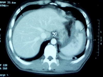
A total of 13 of 23 patients who underwent laparotomy were found to have a bowel or mesenteric injury. Five had bowel injury, 4 had mesenteric hemorrhage, and 4 had both. Mesenteric contrast extravasation was seen in 7 patients, and this correlated with mesenteric bleeding at laparotomy.

The authors found that the following signs on CT scan indicated injury:

  • Full or partial thickness change in bowel wall appearance
  • Increased mesenteric density
  • Free fluid without solid organ injury

Bottom line: This study shows that CT scan can detect bowel and mesenteric injury reliably if you scan the patient 3 times! This seems like over-radiation and overkill. A more intelligent way to approach this would be to perform a normal trauma abdominal scan. If a suspicious area of mesenteric or bowel thickening is seen, then a limited rescan through the affected area only for equilibrium phase images may be warranted. If actual contrast extrvasation is seen, no further scanning is needed. A quick trip to the OR is in order.

Reference: Contrast-enhanced multiphasic computed tomography for identifying life-threatening mesenteric hemorrhage and transmural bowel injuries. J Trauma 71(3):543-548, 2011.

Best Of: IV Contrast

We use CT scanning in trauma care so much that we tend to take it (and its safety) for granted. I’ve written quite a bit about thoughtful use of radiographic studies to achieve a reasonable patient exposure to xrays. But another thing to think about is the use of IV contrast.

IV contrast is a hyperosmolar solution that contains some substance (usually an iodine compound) that is radiopaque to some degree. It has been shown to have a significant impact on short-term kidney function and in some cases can cause renal failure.

Here are some facts you need to know:

  • Contrast nephrotoxicity is defined as a 25% increase in serum creatinine, usually within the first 3 days after administration
  • There is usually normal urine output and minimal to no proteinuria
  • In most cases, renal function returns to normal after 3-4 days
  • Nephrotoxicity almost never occurs in people with normal baseline kidney function
  • Large or repeated doses given within 72 hours greatly increase risk for toxicity
  • Old age and pre-existing diabetic renal impairment also greatly increase risk

If you must give contrast to a patient who is at risk, make sure they are volume expanded (tough in trauma patients), or consider giving acetylcysteine or using isosmolar contrast (controversial, may still cause toxicity).

Bottom line: If you are considering contrast CT, try to get a history to see if the patient is at risk for nephrotoxicity. Also consider all of the studies that will be needed and try to consolidate your contrast dosing. For example, you can get CT chest/abdomen/pelvis and CT angio of the neck with one contrast bolus. Consider low dose contrast injection if the patient needs formal angiographic studies in the IR suite. Always think about the global needs of your patient and plan accordingly (and safely).

Related posts:

Reference: Contrast media and the kidney. British J Radiol 76:513-518, 2003.

How Long Should We Watch Intracerebral Hemorrhage?

Patients with traumatic brain injury (TBI) severe enough to cause bleeding are usually admitted to the hospital for observation and in many cases, repeat CT scanning. Those with small intracranial hemorrhages (ICH) may experience progression of the bleeding, and a small percentage of cases may need operative intervention (1-3%). Questions we typically face are, how long should we watch for progression, and how often should we scan?

A retrospective cohort study was carried out at UMD-NJ, looking for answers for a specific subset of these patients. Specifically, they had to have a mild blunt TBI (loss of consciousness and/or retrograde amnesia, GCS in the ED of 13-15) and a positive head CT. They classified any type of hemorrhage into or around the brain as positive. 

During a 3 year period, 474 adults were enrolled but only 341 were eligible for the study. They were excluded due to previous injury, presence of a mass (not trauma), need for immediate neurosurgical intervention, or failure to get a second CT scan. The authors found:

  • 7% of patients were taking anticoagulants! This is surprisingly high. Interestingly, 15 were subtherapeutic, 3 were therapeutic and 2 were supratherapeutic.
  • Subarachnoid hemorrhage was the most common finding on CT (54%). Intraparenchymal hemorrhage was next most common (48%) Many patients had more than one type of bleed.
  • The injury worsened between the first and second scans in 31% of patients. This number increased to 46% in patients taking anticoagulants. 
  • About 97% of bleeds stopped progressing by 24 hrs post-injury.

Bottom line: Most centers are probably overdoing the observation and repeat scan thing. More than two thirds of bleeds are stable by the first scan (first and second scans identical), and nearly all stop progressing within 24 hours. It’s very likely that patients who are not on anticoagulants and who have a stable neuro exam and stable symptoms can get just one scan and 24 hours of observation. Persistent headache, nausea, failure to ambulate well, or other symptoms warrant a repeat scan and longer observation.

Related posts:

Reference: The temporal course of intracranial haemorrhage progression: How long is observation necessary? Injury 43(12):2122-2125, 2012.

CT Scans And Rib Fractures

Last week I discussed the importance of treating rib fractures in older patients with the greatest respect. One reader commented:

“number of rib fratures are not that accurate by x-ray. If further evaluate by CT, more fractures will be identified”

Well, I agree and I disagree. Chest xray is notoriously inaccurate when it comes to diagnosing or counting rib fractures. Some older studies have shown that a plain chest xray may miss as many as 50% of all rib fractures. On the other hand, CT scan is very accurate at diagnosing them.

But the question is, do we need to know exactly how many ribs are fractured? In general, the answer is no. Rib fracture is a clinical diagnosis. A patient with an appropriate mechanism and focal tenderness on the chest wall has a rib fracture unless proven otherwise. Do we need to prove otherwise? No. They still have pain, and it still needs to be treated. The degree of pain and pulmonary impairment determines the need for admission and more advanced therapies, not an exact count of ribs fractured. 

Bottom line: Rib fracture is a clinical diagnosis! CT scan of the chest for diagnosing rib fractures (or pneumothorax, or hemothorax for that matter) is basically not indicated. It delivers a lot of radiation (and IV contrast if you mistakenly order it), but does not change management. For blunt trauma, CT of the chest should only be used for screening for aortic injury. The only possible indication I can think of is to plan ORIF of complicated, displaced rib fractures. But in that case, let your surgical specialist decide if the test is really necessary.

Related posts:

Undergrading Spleen Injury

We love our CT scans! They’re so high tech, with such detailed images popping up on the monitor so quickly. To take advantage of the detail, we’ve come up with fancy grading systems that can be used to direct care. But are they all they’re cracked up to be?

CT grading of spleen injury is a prime example. We’ve got a nice, detailed system that looks at laceration depth, subcapsular hematoma size and vascular injury. We can use it to predict the likelihood of needing an operation and where we should admit someone in the hospital (ICU vs ward). And when we see the injury on the screen, we believe that we can accurately apply the scoring system to these beautiful images.

But unfortunately, it’s not that simple.Scanning obtains multiple images in an axial plane and lays them out for us to look at. However, the spleen (and most other organs) and not shaped like a cube. It is curved, with complex nooks and crannies that can look like cracks. Moderate to large hematoma around the spleen can obscure lacerations. And the hilum is even more complicated and variable in shape.

Because of this, CT scans of the abdomen tend to underestimate the true extent of injury, especially in the higher grades. Grade I and II injuries are usually accurate, but in Grades III-V, the scan tends to undergrade by 1 (30% of cases) or 2 grades (45% of cases) when re-graded at surgery.

Bottom line: Grade I and II injuries are generally managed in a lower intensity setting and almost never require operation. But beware of the higher grades! It is very likely that it’s higher than you think. This means that if your patient slowly becomes tachycardic or their blood pressure softens, believe the clinical evidence. Don’t rely on a CT scan that was done hours ago that may be hiding a more severe injury than you think! (This applies to liver injuries as well)

Related posts:

Reference: Correlation of operative and pathological injury grade with computed tomographic grade in the failed nonoperative management of blunt splenic trauma. Euro J Trauma Emerg Surg – Online First 2 Mar 2012.