Tag Archives: coagulopathy

Best of EAST 2024 #2: Prehospital End Tidal CO2 And Fibrinolysis

Coagulopathy is the bane of every trauma professional. Trauma patients are bleeding to death until proven otherwise, and once they start bleeding, it only gets worse. A key component of this issue is the presence of fibrinolysis, which commonly occurs after severe trauma. Although the prime objective in managing these patients is definitive control of bleeding, antifibrinolytic therapy such as tranexamic acid (TXA) may be beneficial during the time before that can happen.

The trauma group at Denver Health has been studying fibrinolysis and finding things to do with TEG machines for many years. They postulated that, since hemorrhagic shock can cause hyperfibrinolysis (HF) and early administration of agents like TXA seem to work better when given early, wouldn’t it be nice to have a more objective way of identifying it as early as possible?

They designed a prehospital study that used end-tidal CO2 monitoring in the ambulance and correlated results with a TEG reading upon arrival at the hospital.  The study was prospective and observational and involved two Level I trauma centers. End-tidal CO2 was measured from the ventilator circuit or via nasal cannula capnography. The authors compared this reading to other possible shock indicators, such as systolic blood pressure and the shock index.

Here are the factoids:

  • A total of 138 patients were studied, and 13 had hyperfibrinolysis identified on hospital arrival
  • Of the 13, 9 required massive transfusion, and eight died
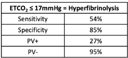
  • An ETCO2 value <17 mm Hg was determined to have a positive predictive value of 27% and a negative predictive value of 95%
  • The area under the receiver operating characteristic curve was 0.71, which was better than the blood pressure (0.58) and shock index (0.54)

The authors concluded that the ETCO2 was an accurate, objective, inexpensive, and noninvasive method of measuring the risk of hyperfibrinolysis that could guide the use of agents such as TXA.

Bottom line: A lot is going on in this abstract. The central concept is that it is trying to identify a surrogate for TEG-identified fibrinolysis available in the field. It compares ETCO2 with two other semi-objective indicators, blood pressure and shock index (pulse divided by blood pressure).

The biggest issue is that the number of patients with fibrinolysis was very small, only 13. Statistical comparisons of variables between the two groups are difficult because the number of HF patients in several subgroups was only 4 or 5. 

The sensitivity, specificity, and positive/negative predictive values are so-so. If the ETCO2 is above the 17mm Hg threshold, the likelihood of patients not having HF is good at 95%. But if it is below, the likelihood that they actually have HF is only 27% (true positive rate).
The area under the curve calculations is also not very impressive. Yes, an AUC of 0.71 is better than 0.54-0.58, but it is still not great.

One always has to be careful finding surrogates (ETCO2) for things you really want to measure (TEG LY30 > 3%). Many potential confounders can limit their usefulness. And this case is no different, which should be apparent from the numbers. Perhaps the data would be better if a much larger group of patients were studied. Unfortunately, this will probably take close to 1,000 subjects and require a multicenter trial. 

This is interesting preliminary work. It’s definitely not enough to change practice now. But with more work, and more patients, who knows?

Reference: Prehospital ETCO2 predicts hyperfibrinolysis in injured patients: implications for early use of antifibrinolytics in trauma. EAST 2024 Podium paper 3.

Hypothermia and Massive Transfusion

Tuesday, I talked about a new notion of using profound hypothermia to save critically injured trauma patients. Since this concept is not yet ready for prime time, we still have to treat hypothermia as our enemy. Most trauma centers have established massive transfusion protocols that detail the use and ratios of specific blood components to avoid fatal anemia and coagulopathy. But do we pay enough attention to hypothermia?

A multicenter study was carried out that will be reported at the upcoming EAST meeting in January. They looked at patients who received massive transfusion (>= 10u PRBC in 24 hours) and looked at their lowest temperature during that 24 hour period. 

They found that as temperature decreased, shock parameters, coagulopathy, injury severity and transfusion requirements increased significantly. Specifically, if a temperature of <34C doubled mortality risk, and this effect was most pronounced in patients who received relatively less plasma.

Bottom line: Temperature is still very important, and hypothermia must be avoided at all costs. This is true in the ED and the OR. Allowing temperature to drop below 34C significantly increases mortality and is at least as important as giving enough FFP to correct coagulopathy from dilution.

Related post:

Reference: Hypothermia in massive transfusion: are we not paying enough attention to it? Poster 2, EAST 25th Annual Assembly, Jan 2012.

What’s The INR of Fresh Frozen Plasma?

So what’s the INR of FFP? Or stated another way, what’s the lowest you can correct a patient’s INR using infusions of fresh frozen plasma?

One of the mainstays of correcting coagulopathy, either from hemorrhage or due to medication like warfarin, is transfusion of FFP. Frequently, clinicians will write orders to administer FFP until a certain INR is achieved. What is a reaonable INR?

A “normal” INR is 1.0, plus or minus about 0.2, depending on your laboratory. However, two separate studies have shown that transfusion of FFP will not reliably decrease the INR below about 1.7. 

Bottom line: The answer to the question is about 1.6. If any clinician orders FFP transfusions with a goal INR below this, it probably won’t happen. And since transfusions of any product have risks, my “juice to squeeze” ratio of risk vs benefit begins to fail at an INR of 1.6. Below that point, the patient needs a normal temperature and good perfusion to drop their INR further.

References:

  • Toward rational fresh frozen plasma transfusion: the effect of plasma transfusion on coagulation test results. Am J Clin Pathol 126(1):133-139, 2006.
  • Effect of fresh frozen plasma transfusion on prothrombin time and bleeding in patients with mild coagulation abnormalities. Transfusion 46(8):1279-1285, 2006.

Early Coagulopathy After Severe Traumatic Brain Injury Equals Poor Outcome

Coagulopathy is a frequent occurrence after severe traumatic brain injury (TBI). There are high levels of tissue factor (TF) in the brain, which can be released with severe injury. This in turn triggers a cascade which can lead to generalized coagulopathy.

The trauma group at LAC+USC looked at the time course of coagulopathy after isolated severe TBI. They identified 278 patients over a 1.5 year period and retrospectively review a number of demographic and outcome variables. Coagulopathy was defined as a platelet count < 100,000/mm3, INR > 1.4, or PTT > 36 sec.

They found the following:

  • 46% with blunt trauma and 82% with penetrating injury developed a coagulopathy 
  • Presence of coagulopathy increased with increasing head injury severity
  • Thromobocytopenia as a cause of coagulopathy was less common (17%) than clotting factor problems
  • As brain injury severity increased from AIS=3 to AIS=5, median onset of coagulopathy became increasingly earlier (26 hrs, 22 hrs, 10 hrs)
  • Mortality increased with earlier coagulopathy (23% after 24 hrs, 39% between 12 and 24 hrs, 56% less than 12 hrs)

Bottom line:

  • Prehospital: Coagulopathy should be suspected if the patient is bleeding profusely from multiple sites, including your IV needle sticks. This indicates severe brain injury and demands triage to a trauma center with immediate neurosurgical support.
  • In-hospital: Coagulopathy that is noted in the ED portends severe injury and poor prognosis. Rapid access to CT scan and your neurosurgical consultant is critical.

Related post: Controlling fever in head injury

Reference: Time course of coagulopathy in isolated severe traumatic brain injury. Injury 41:924-928, 2010.

Evolution of Use of Recombinant Factor VIIa

Recombinant Factor VIIa was initially approved for bleeding in hemophiliac patients back in 1999. Over the years, there has been a big move toward off-label use. There appeared to be obvious utility in using it as an emergency hemostatic agent in trauma patients. But as with many new drugs and devices, early enthusiasm slowly gave way to more balanced judgment. Reviews during the past few years are less glowing than they were early on. So what’s really been happening over the past decade?

Researchers at Stanford tapped into a large database of patient level records from 600 hospitals around the US. They identified over 18,000 uses of Factor VIIa during a 9 year time period. By the end of the study period (2008), 97% of use was off-label! Approved use (hemophilia) increased 4-fold, while off-label use increased by 140-fold. Cardiovascular surgery and trauma tied in their amount of off-label use (both about 29% of the sample).

Does it do any good? This paper can’t directly address that question, since it does not have a good comparison group. However, looking at in-hospital mortality is revealing. Use for hemophilia (FDA approved) results in a 4% mortality rate. For trauma, the in-house mortality is 33%. The worst outcomes were with patients with an aortic aneurysm (55% mortality). 

Bottom line: This review details the administration of about $175 million worth of recombinant Factor VIIa over 9 years. Off-label use has skyrocketed despite a dearth of good reports that it actually saves lives. The Number Needed to Treat to prevent one additional bad outcome keeps getting larger with every study published. With a price tag of nearly $10,000 per dose, it’s getting harder to justify using it. I think we are seeing the beginning of the end (at least in trauma) for this powerful drug.

Reference: Off-label use of recombinant Factor VIIa in US hospitals: analysis of hospital records. Annals of Int Med 154(8):516-522, 2011.