Tag Archives: Cervical spine

Trauma Mythbusters: Spinal Cord Injury From Airway Management

Airway management is one of the most anxiety provoking procedures performed by trauma professionals. The main fear is that the airway will be lost during attempts to secure it and patient demise will soon follow. Add some facial fractures, bleeding, and an “unfavorable body habitus”, and the average prehospital or ED provider is really on edge.

The next most common fear is that providing a definitive airway in a patient with a known (or even suspected) cervical spine injury could cause a catastrophic neurologic injury. This was first addressed back in 1989 (before the time of video laryngoscopy, and when flexible scopes were rarely available in the ED). The authors found no verifiable reports of such an injury in the entire English literature

Over the years, a few case reports have cropped up. As in so much of the medical literature, causality is hard to prove. The patient was normal before anesthesia, and afterwards they were not. Had to be the intubation, right?

Not so fast! Let’s break it down and look at what we do know. Biomechanical studies have shown that the manipulation that occurs in direct laryngoscopy isn’t as bad as it looks. Studies in uninjured models are enlightening (minimal movement with blade insertion, slight rotational movements with blade elevation, and a little more rotation during the intubation). Most of this (slight) movement occurs from occiput to C2, with little motion at all at C3 and below.

But that was on an uninjured model. What about ones that simulate an injured spine? Specifically an injury in the upper spine area that we know moves?

  • Cricoid pressure caused no appreciable changes in the spine
  • Chin lift and jaw thrust reduced space available for the cord (SAC) by 1 and 2.5mm, respectively, and caused an angulation of about 4-5 degrees
  • SAC narrowed by only 1.5mm, even with maximum flexion and extension
  • Oral and nasal intubation narrowed SAC by 1.6mm, and resulted in a maximum of 2.5 degrees of rotation
  • Video laryngoscopy results in about half of the rotational movement of direct laryngoscopy
But what about these sporadic reports of neurologic deficits after intubation? What is often neglected is that spinal blood flow and long-term neck positioning have a major impact on cord function. Even relatively mild malpositioning of the cervical spine for extended periods during an OR case have been documented. 

Bottom line: From a mechanical standpoint, even in unstable spine models, the maneuvers we use in preparation for intubation cause more movement of the spine than does the intubation procedure itself. The true number of spinal cord injuries actually (and provably) caused by intubation approaches zero. The literature suggests that video laryngoscopy results in less overall movement during intubation, but it doesn’t seem to have an impact on cord injury (you can’t get less than zero). 

References:

  • Spinal cord injury and direct laryngoscopy – the legend lives on. Br J Anesth 84(6):705-709.
  • Airway management in adults after cervical spine trauma. Anesthesiology 104(6):1293-1318, 2006.

How To: Flexion / Extension Views of the Cervical Spine

Clearance of the cervical spine can often be done using clinical criteria alone (see this video at http://youtu.be/NhjF9kDOcjE). If this is not possible, a combination of radiologic and clinical evaluation is usually carried out. 

In some cases, radiographic studies (usually CT) are normal, but there is pain on clinical exam. Our next step is to send the patient to xray for flexion and extension views. This exam is performed by removing the collar while the patient is sitting, so the thoracic and lumbar spines must be clear before ordering this. The patient then gently flexes and extends the neck to their limits of comfort. Images are then obtained at the limits of flexion and extension. The premise is that a normal, awake patient cannot and will not move their neck beyond their comfort level to the point where they could cause themselves neurologic injury. 

It is very important that you look at the images yourself. The radiologist may review the images and will report that “there is no evidence of subluxation at the limits of flexion and extension.” But the patient may have barely moved their neck!

The question is: how much flexion and extension do you need to have to clear the spine?

The answer is not easy to find, and is buried in literature from the 1980s and 90s. According to the EAST guidelines, the ideal amount is 30 degrees from neutral for both flexion and extension. This is not always achievable in elderly patients, so in those cases you must use your judgment. Talk to the patient to find out if they stopped moving their neck forward or backward due to pain, or because they just can’t move it that far.

Trouble signs to look for are:

  • Subluxation of more that 2mm at any level
  • Angulation of more than 11 degrees

Any abnormality should prompt a spine consult.

If the study is not abnormal but the amount of flexion and/or extension is not adequate, there are two options. First, just leave the collar in place and try again in a week or so and try again. This will allow any soft tissue injuries to get better and may allow a successful repeat study. The alternative is a more costly and less well-tolerated MRI.

References:

  1. EAST Practice Guidelines, Identifying Cervical Spine Injuries Following Trauma – Update (2000).
  2. Defining radiographic criteria for flexion-extension studies of the cervical spine. Robert Knopp et al. Ann Emerg Med. 2001 Jul;38(1):31-5.

Is Applying Or Removing That Cervical Collar Dangerous?

Cervical collars are applied to blunt trauma patients all the time. And most of the time, the neck is fine. It’s just those few patients that have fracture or ligamentous injury that really need it. 

I’ve previously written about how good some of the various types of immobilization are at limiting movement (click here). But what happens when you are actually putting them on or taking them off? Could there be dangerous amounts of movement then?

Several orthopaedics departments studied this issue using an electromagnetic motion detector on “fresh, lightly embalmed cadavers” (!) to determine how much movement occurred when applying and removing 1- and 2-piece collars. Specifically, they used an Aspen 2-piece collar, and an Ambu 1-piece. They were able to measure flexion/extension, rotation and lateral bending.

There were no significant differences in rotation (2 degrees) and lateral bending (3 degrees) when applying either collar type or removing them (both about 1 degree). There was a significant difference (of 0.8 degrees) in flexion/extension between the two types (2-piece flexed more). Movement was similarly small and not significantly different in either collar when removing them.

Bottom line: Movement in any plane is less than 3-4 degrees with either a 1-piece or 2-piece collar. This is probably not clinically significant at all. Just look at my related post below, which showed that once your patient is in the rigid collar, they can still flex (8 degrees), rotate (2 degrees) and move laterally (18 degrees) quite a bit! So be careful when using any collar, but don’t worry about doing damage if you use it correctly.

Related post:

Reference: Motion generated in the unstable cervical spine during the application and removal of cervical immobilization collars. J Trauma 72(6):1609-1613, 2012.

Best Of: Flexion / Extension Views of the Cervical Spine

I’ve gotten a number of requests about the use of flexion-extension views of the cervical spine to aid in spine clearance. Here are some answers to common questions about this practice.

Clearance of the cervical spine can often be done using clinical criteria alone (see this video at http://youtu.be/NhjF9kDOcjE). If this is not possible, a combination of radiologic and clinical evaluation is usually carried out. 

In some cases, radiographic studies (usually CT) are normal, but there is pain on clinical exam. Our next step is to send the patient to xray for flexion and extension views. This exam is performed by removing the collar while the patient is sitting, so the thoracic and lumbar spines must be clear before ordering this. The patient then gently flexes and extends the neck to their limits of comfort. Images are then obtained at the limits of flexion and extension. The premise is that a normal, awake patient cannot and will not move their neck beyond their comfort level to the point where they could cause themselves neurologic injury. 

It is very important that you look at the images yourself. The radiologist may review the images and will report that “there is no evidence of subluxation at the limits of flexion and extension.” But the patient may have barely moved their neck!

The question is: how much flexion and extension do you need to have to clear the spine?

The answer is not easy to find, and is buried in literature from the 1980s and 90s. According to the EAST guidelines, the ideal amount is 30 degrees from neutral for both flexion and extension. This is not always achievable in elderly patients, so in those cases you must use your judgment. Talk to the patient to find out if they stopped moving their neck forward or backward due to pain, or because they just can’t move it that far.

Trouble signs to look for are:

  • Subluxation of more that 2mm at any level
  • Angulation of more than 11 degrees

Any abnormality should prompt a spine consult.

If the study is not abnormal but the amount of flexion and/or extension is not adequate, there are two options. First, just leave the collar in place and try again in a week or so and try again. This will allow any soft tissue injuries to get better and may allow a successful repeat study. The alternative is a more costly and less well-tolerated MRI.

Related posts:

References:

  1. EAST Practice Guidelines, Identifying Cervical Spine Injuries Following Trauma – Update (2000).
  2. Defining radiographic criteria for flexion-extension studies of the cervical spine. Robert Knopp et al. Ann Emerg Med. 2001 Jul;38(1):31-5.
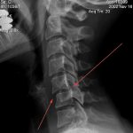
Image: C5-6 subluxation with only a slight amount of flexion