Category Archives: Thorax

What’s Wrong With My Patient? Final Answer!

I previously described a young man who was recovering from surgery for repair of a stab to the heart. He presented shortly after discharge with fever, a slightly elevated WBC, some EKG changes, and a small pericardial effusion.

Several people tweeted out the answer, which is post-pericardiotomy syndrome (PPS).

PPS is an inflammatory reaction to traumatic and then surgical injury to the pericardium. It is seen in a relatively small percentage of patients who undergo pericardiotomy for trauma, which is why patients who develop it are such a surprise to trauma professionals. A similar condition can develop after myocardial infarction (Dressler syndrome) and was first described in 1956. The classic paper describing PPS after trauma was published five years later (cited below).

Symptoms typically develop 1-6 weeks after surgery, and usually consist of low grade fevers, malaise, chest pain, and occasionally arthralgia. A pericardial friction rub may be present (and where is that stethoscope, BTW?). The WBC is usually elevated, with some degree of left shift. EKG may show some degree of pericarditis, including global ST elevation and T wave inversion.

Chest x-ray is usually nonspecific, but may show pleural effusion or an enlarged heart due to the presence of some pericardial fluid. Ultrasound may confirm a pericardial effusion, but this is not a reliable finding since the pericardium is typically left open at the end of the operation.

Treatment is symptomatic, usually consisting of NSAIDS or aspirin to tone down the inflammatory response.  These drugs are usually given for 4-6 weeks, then tapered. If the effusion is large, pericardiocentesis may be needed.

Bottom line: If your postop heart injury patient presents with these symptoms, consider infectious etiologies first, but remember that they are typically even less common than post-pericardiotomy syndrome. Reassure your patient, then reach for the ibuprofen to get them through it.

Reference: Postpericardiotomy syndrome following traumatic hemopericardium. Am J Cardiology 7(1):83-96, 1961.

What’s Wrong With My Patient? Part 2

In my previous post, I described a young man who had recovered from a stab to the heart. He did well for a week and a half, but then presented to the ED with significant chest pain. It seems to be substernal and somewhat pleuritic. What should you do to work it up further?

There have been a number of helpful comments. The first order of business is to rule out problems which may prove to be life threatening. In his case, ischemic disease and some failure of the repair must be ruled out quickly. Although ischemia or MI are unlikely in this young man, they are possible and should be evaluated.

I recommend the following:

  • Auscultate the chest and heart (remember this from medical school?)
  • PA chest x-ray
  • EKG
  • CBC
  • Troponin
  • FAST exam focusing on the heart

My list is short and simple, and should help me figure out nearly all significant problems.

In this case, the following findings are present:

  • The lungs are clear, and their is a faint cardiac friction rub
  • The chest x-ray is unremarkable
  • EKG shows ST elevations in two of the lateral leads only. Otherwise, it is normal.
  • CBC is normal with the exception of WBC 14,000
  • There is a trace level of troponin present
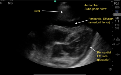
  • FAST demonstrates a very small pericardial effusion without clot

So what do you make of all this? What’s the diagnosis? What do you need to do? Tweets and comments please.

Answers Monday!

What’s Wrong With My Patient?

I’ve had several requests for this case recently, so I figured I’d put it out there again.

A 25 year old man is involved in some sort of violent, non-productive interpersonal relationship. He sustains a stab to the left chest, and is brought to your trauma center as a trauma team activation. During the FAST exam, a moderate effusion with visible clot is seen in the pericardium.

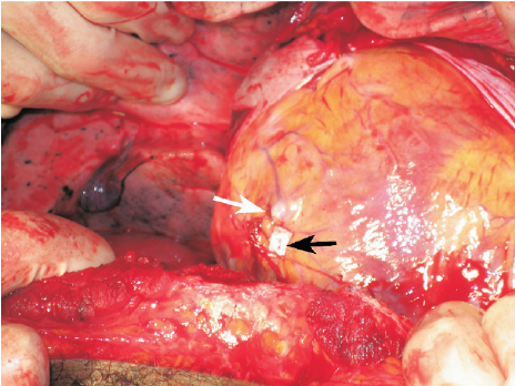
Appropriately, you run to the OR and prepare for a left thoracotomy. You perform a pledgeted repair of the ventricle (black arrow) and close.

The patient does well and is discharged home five days later. He returns to your clinic the following week and is doing well. You remove the staples.

One week later, he returns to your emergency department complaining of significant chest pain. He describes it as deep, behind his sternum, and it seems to be exacerbated by breathing.

Now what? What are you thinking about? What additional exam do you need. What labs?

Tweet or comment with your answers and suggestions. More on this tomorrow!

Flying Or Diving After Traumatic Pneumothorax

Patients who have sustained a traumatic pneumothorax occasionally ask how soon they can fly in an airplane or scuba dive after they are discharged. What’s the right answer?

The basic problem has to do with Boyle’s Law (remember that from high school?). The volume of a gas varies inversely with the barometric pressure. So the lower the pressure, the larger a volume of gas becomes. Most of us hang out pretty close to sea level, so this is not an issue. But for flyers or divers, it may be.

Flying

Helicopters typically fly only one to two thousand feet above the ground, so the air pressure is about the same as standing on the earth. However, flying in a commercial airliner is different. Even though the aircraft may cruise at 30,000+ feet, the inside of the cabin remains considerably lower though not at sea level. Typically, the cabin altitude goes up to about 8,000 to 9,000 feet. Using Boyle’s law, any volume of gas (say, a pneumothorax in your chest), will increase by about a third on a commercial flight.

The physiologic effect of this increase depends upon the patient. If they are young and fit, they may never know anything is happening. But if they are elderly and/or have a limited pulmonary reserve, it may compromise enough lung function to make them symptomatic. And having a medical problem in an aluminum tube at 30,000 feet is never good.

Commercial guidelines for travel after pneumothorax range from 2-6 weeks. The Aerospace Medical Association published guidelines that state that 2-3 weeks is acceptable. The Orlando Regional Medical Center reviewed the literature and devised a practice guideline that has a single Level 2 recommendation that commercial air travel is safe 2 weeks after resolution of the pneumothorax, and that a chest x-ray should be obtained immediately prior to travel to confirm resolution.

Diving

Diving would seem to be pretty safe, right? Any pneumothorax would just shrink while the diver was at depth, then re-expand to the original size when he or she surfaces, right?

Not so fast. You are forgetting why the pneumothorax was there in the first place. The lung was injured, most likely via tearing it, penetration by something sharp, or popping a bleb. If the injured area has not completely healed, then air may begin to escape through it again. And since the air used in scuba diving is delivered under pressure, this could result in a tension pneumothorax.  This is disastrous underwater!

Most injuries leading to pneumothorax heal completely. However, if there are bone spicules stuck in the lung or more complicated parenchymal injuries from penetrating injury, they may never completely heal. This makes the diver susceptible to a tension pneumothorax anytime they use their regulator.

Bottom line: Most patients can safely travel on commercial aircraft 2 weeks after resolution of pneumothorax. Ideally, a chest xray should be obtained shortly before travel to confirm that it is gone. Helicopter travel is okay at any time, since they typically fly at 1,500 feet or less.

Divers should see a physician trained in dive medicine to evaluate their injury and imaging prior to making another dive.

References:

  • Divers Alert Network – Pneumothorax – click to download
  • Practice Guideline, Orlando Regional Medical Center. Air travel following traumatic pneumothorax. October 2009.
  • Medical Guidelines for Airline Travel, 2nd edition. Aerospace Medical Association. Aviation, Space, and Environmental Medicine 74(5) Section II Supplement, May 2003.