Category Archives: Procedures

ED Thoracotomy Survey: Read The Answers! (US)

Again, thanks for all who submitted their survey answers. Here’s a rundown of the answers provided by US respondents. A few duplicates from the same hospitals have been merged into single answers for them. Total number of US centers for the tables below is 149.

Level of trauma center

Level I 83
Level II 37
Level III 15
Level IV 1
Level V 2
Seeking verification/designation 1
No level 10


How many ED thoracotomies are performed per year at your hospital?

A few per year (<6) 83
About every month (6-15) 35
A couple of times a month (16-30) 23
About every week (31-52) 8


What type of trauma do you perform ED thoracotomy for?

Both blunt and penetrating 79
Penetrating 64
Blunt 5


Do you use a practice guideline for ED thoracotomy?

Yes 86
No 47
I’m not sure 15


Do you use REBOA in your ED?

No 88
Yes 58
I’m not sure 3


And now for the questions you’ve been waiting for!

Who could perform ED thoracotomy at your hospital? (n=149)

Surgeon 145
Emergency physician 109
Surgical resident / fellow 93
Emergency medicine resident 66
APP (PA, NP) 2 at one Level I and one Level V
Family physician 1 at one Level V
Family medicine resident 1 at one Level V


Who usually performs ED thoracotomy at your hospital? (n=149)

Surgeon 115
Emergency physician 25
Surgical resident / fellow 69
Emergency medicine resident 17
Never done one 3
Family physician or family nurse practitioner 1 at one Level V


Who usually performs ED thoracotomy at your hospital? (By trauma center level)

Level I (n=83) II (n=37) III (n=15)
Surgeon 64 35 11
Emergency physician 8 3 6
Surgical resident 63 4 1
Emergency medicine resident 12 1 2
No one 0 0 1


Join me tomorrow when I review the international data!

Print Friendly, PDF & Email

Coming Tomorrow: ED Thoracotomy Survey Results

The data is in!

Thanks to everybody (all 200+ of you) who participated in the ED thoracotomy survey over the past month. I’m currently compiling the results and will post them here over the next two days.

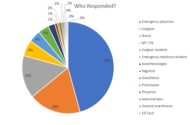
Here is a summary of who responded:

  • 50% were emergency medicine physicians or residents
  • 22% were surgeons or surgical residents
  • 15% were nurses
  • 6% were advanced practice providers such as NPs or PAs

And where were they located?

As you can see, the vast majority (167) were from the United States. Australia, Canada, and Denmark added another 18, and a variety of other countries contributed the remaining 27 surveys.

Over the next two days, I’ll focus on the US data, then look at the results from the rest of the world.

Again, thanks for contributing!


Print Friendly, PDF & Email

Long-Term Experience With Endovascular Aortic Repair For Trauma

For decades, the treatment of blunt injury to the thoracic aorta was open repair. The big debate at the time was use of cardiac bypass vs fast clamp and sew. But starting in 1997 with the introduction of thoracic endovascular aortic repair (TEVAR) of this injury, we have rapidly moved to the point where most traumatic aortic injuries are repaired using this technique.

A report that was written nearly a decade ago indicated a relatively high complication rate for the procedure. Graft complications were reported in 18% of patients, with 14% showing endoleaks. Stroke and left arm ischemia were also reported.

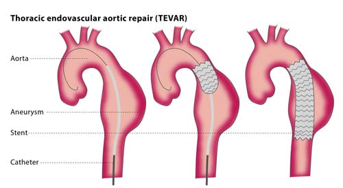
The diagram above shows insertion for management of an aneurysm, but the technique is similar for trauma. Blunt aortic injury occurs closer to the left subclavian artery and care must be taken to place the endograft closer to but not covering its orifice.

As the insertion systems and stents improved, short term events have been on the decline. Unfortunately, long term followup data has been hard to come by.

Until now. An article that is not yet in print reports 11 years of experience and followup with patient undergoing TEVAR at the ShockTrauma center in Baltimore.

Here are the factoids:

  • 88 patients underwent TEVAR during the study period, all from blunt trauma
  • Average ISS was 38, showing these patients were severely injured
  • Overall mortality was 7%, but none was due to the TEVAR procedure
  • TEVAR-related complication rate was 9% Endoleaks at the ends of the graft occurred in 4 patients, and all required repair. There were 4 other minor leaks that resolved on their own.
  • 26 had all or part of the left subclavian orifice covered at initial operation. None developed ischemia, although 2 had a prophylactic carotid-subclavian bypass before TEVAR.
  • The longest followup imaging occurred 8 years after the procedure. No long-term complications were noted.

Bottom line: TEVAR has essentially replaced open repair of the aorta, except in special cases. We continue to learn from our experience, and the complication rate is still falling. Other than endoleaks recognized in the postop period, most other complications rarely occur. Long term followup is poor, but in the patients who do return, there were no complications. But remember, this is an expected sampling bias. If the patient had major problems and/or died, they would just be lost to followup. We would never know.

Print Friendly, PDF & Email

EAST 2017 #10: A Simple Way To Predict Complications After Rib Fracture?

Rib fractures are a common injury, and a very common cause of morbidity. Every time I admit an elderly patient with rib fractures, I debate whether they should go to the ICU or a ward bed. Could there be a more objective way of determining the likelihood of complications, aggressiveness of treatment, and admission unit?

A group at West Virginia University implemented a rib fracture pathway in 2009, and have been collecting data on patients ever since. It was based on the measurement of forced vital capacity (FVC) on admission. This is the total amount of air that can be exhaled during a forced breath.

The authors subdivided their patients into two groups based on the total volume exhaled (<1.5L, and >1.5L). They retrospectively reviewed 6 years of data, looking at specific injuries, complications, and unexpected transfer to ICU. They hypothesized that patients in the highest FVC group would have fewer complications.

Here are the factoids:

  • There was a nearly even split in groups, with 678 patients who had FVC > 1.5L, and 682 with FVC < 1.5
  • There were significantly fewer complications and pneumonia, as well as fewer readmissions in the FVC > 1.5 group
  • Higher FVC was not associated with fewer unexpected transfers to ICU
  • Length of stay was half as long (4d vs 8d) in the high FVC group, but no p value was provided
  • The authors conclude that patients with FVC much greater than 1.5 are at lower risk for complications regardless of the number of fractures (???!)
  • They even suggest that patients with FVC > 1.5 could be discharged from the ED rather than be admitted (!)

Bottom line: Well, it started out good! The abstract showed that the high FVC patients had fewer complications and readmissions. And the length of stay was shorter, although significance was not noted. But the jump to correlating complication risk with number of fractures was not addressed in the abstract. And I can’t quite grasp the leap to suggesting possible discharge from the ED. 

FVC may be an inexpensive and simple test to administer in new rib fracture patients. But it’s ability to predict who goes to ICU and who goes home from the ED was not really identified in the study. 

Questions and comments for the authors/presenters:

  1. A minor point, but the upper limit was defined as > 1.5L in some parts of the abstract, and > 1.5L in  others. Small point, but keep it clean. Make sure all the greater than, less than, and equals signs are consistent.
  2. Was the shorter length of stay significantly different between the groups?
  3. Did you do any stratification by age?
  4. How did you make the conclusion that patients could be sent home from the ED?
  5. And did you do any correlations with your FVC data and the number of fractures? It’s not in the abstract.

Click here to go the the EAST 2017 page to see comments on other abstracts.

Related post:

Reference: Is an FVC of 1.5 adequate for predicting respiratory sufficiency in rib fractures? Paper #4, EAST 2017.

Print Friendly, PDF & Email

EAST 2017 #7: Pigtail vs Chest Tube – Does Size Matter?

I’ve been somewhat old school when it comes to chest tubes. Unlike some, I don’t believe that you have any control of where a chest tube goes if you are placing it in a closed chest. Only in the OR with an open one. And I’ve got plenty of x-rays to prove it.

And I used to think that chest tube size mattered when dealing with hemothorax. In theory, you need a big tube to get clots out, right?

Well, maybe not! The trauma group at the University of Arizona Tucson has previously done work on using 14 French pigtail catheters in lieu of a full-size tube. They will be presenting their extended experience with this concept at EAST 2017.

They have maintained a prospectively collected database of information on trauma patients with chest tubes for many years. This study focused only on those who had blood in their chest, either hemothorax (HTX) or hemopneumothorax (HPTX). They also looked at trends in their selection of chest drain tubes.

Here are the factoids:

  • Nearly 500 patients were treated with a tube for HTX or HPTX during the 7 year study period, 2/3 with a chest tube and 1/3 with a pigtail
  • Pigtails had more fluid drain initially (430cc vs 300cc, significant), and 1 less treatment day (4 vs 5, also significant)
  • Failure rate and insertion-related complications were the same (about 22% and 6%, respectively)
  • The group found that their use of pigtails steadily and significantly increased over the years

Bottom line: I’m coming around. The literature does appear to be tilting toward smaller tubes, and this longer-term study helps confirm that. How can this be? Although this is speculation on my part, it probably has to do with the fact that any size tube will drain liquid blood. And probably no size of tube will successfully get all the clot out. 

And certainly, smaller tubes are much better tolerated and do not require the degree of sedation that a mega-tube does. The authors suggest that a multi-center trial should be carried out to confirm this. For my part, I’m going to review the literature we have to date and consider modifying my own chest tube policy (see links below).

Questions and comments for the authors/presenters:

  • Where did you typically insert the pigtails? Anterior chest or classic chest tube position? Was it consistent?
  • Was/is the selection of tube type an attending surgeon specific choice, or did you implement a policy to direct them?
  • Did patient injury pattern or body habitus have any part in tube selection?
  • What about removal failures? That is, how many had to have a tube replaced, and how many went on to require VATS or other surgical procedure for drainage?
  • I enjoyed this provocative paper!

Click here to go the the EAST 2017 page to see comments on other abstracts.

Related posts:

Reference: A prospective study of 7-year experience of using percutaneous 14-French pigtail catheter for traumatic hemothorax at a Level I trauma – size still does not matter. Quick Shot #4, EAST 2017.

Print Friendly, PDF & Email