Category Archives: Procedures

COVID-19 Thinking Cap: How To Protect Personnel During Intubation (Video)

There is a fascinating letter in the New England Journal of Medicine submitted by authors from the Boston Medical Center and Brigham and Women’s Hospital. Like all trauma professionals, they were concerned with droplet contamination produced during the intubation process. Most hospitals have modified their intubation procedures to try to protect personnel as much as possible.

The authors designed a Plexiglas box with two holes for the arms of the intubator that is placed over the patient’s head. This should serve to shield them, and other personnel in the room if the patient unexpectedly coughs during the process. They tested this concept using an intubation mannequin. First, they placed a balloon filled with fluorescent dye in its mouth and slowly inflated until it burst. Here was the result when viewed under ultraviolet light. Sputum everywhere!

Next, they placed the intubation shield over the patient. Here is a drawing of its dimensions.
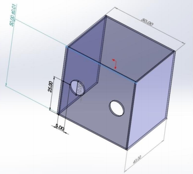
The device is open on the bottom and on the side away from the intubator. The arm holes are 10cm in diameter.

The authors then repeated the balloon experiment with the shield in place and the intubator’s arms inserted through the holes. The resulting contamination was limited to their hands and forearms, and the inside of the shield.

Bottom line: This is a very interesting yet simple and cheap device that can be built by just about anyone and should protect personnel from droplet contamination. It will not have much effect on aerosols escaping into the room, but that’s what our other PPE are for! It’s a great example of how creativity is key in keeping us all safer during this pandemic.

You can view the video on the NEJM website at:
https://www.nejm.org/doi/full/10.1056/NEJMc2007589

Reference: Barrier Enclosure during Endotracheal Intubation. NEJM DOI: 10.1056/NEJMc2007589, April 4 2020.

Air Embolism From an Intraosseous (IO) Line

IO lines are a godsend when we are faced with a patient who desperately needs access but has no veins. The tibia is generally easy to locate and the landmarks for insertion are straightforward. They are so easy to insert and use, we sometimes “set it and forget it”, in the words of infomercial guru Ron Popeil.

But complications are possible. The most common is an insertion “miss”, where the fluid then infuses into the knee joint or soft tissues of the leg. Problems can also arise when the tibia is fractured, leading to leakage into the soft tissues. Infection is extremely rare.

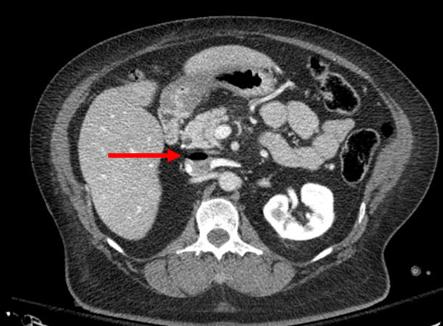
This photo shows the inferior vena cava of a patient with bilateral IO line insertions (black bubble at the top of the round IVC).

During transport, one line was inadvertently disconnected and probably entrained some air. There was no adverse clinical effect, but if the problem is not recognized and the line is not closed properly, there could be.

Bottom line: Treat an IO line as carefully as you would a regular IV. You can give anything through it that can be given via a regular IV: crystalloid, blood, drugs. And even air, so be careful!

Trends In IVC Filter Placement And Retrieval

Yesterday, I reviewed a paper that highlighted a single-institution experience for IVC filter usage. Today, let’s look at a much larger pool of data.

Placement of a filter in the inferior vena cava (IVC) is one of the many tools for managing pulmonary embolism. There was a significant increase in filter placement during the 1990s and 2000s due to a broadening of the indications for its use.  There has been continuing debate over the complications and efficacy of use of this device.

A paper from NYU Langone Health in New York City, the Harvey L. Neiman Health Policy Institute, and Georgia Institute of Technology School of Economics looked a long-term trends in IVC filter use in the Medicare population. They scanned a Centers for Medicare and Medicaid Services (CMS) database over the 22 year period from 1994 to 2015. They specifically analyzed trends in insertion, removal, placement setting, and specialty of the inserting physician.

Here are the factoids:

  • 2008 seemed to be the heyday of IVC filter insertion. Rates nearly tripled by 2008, but have declined about 40% since then (see below). Pay attention to the retrieval rates.

  • Overall, filters were most commonly placed by radiologists, followed by surgeons and cardiologists. Here’s the diagram above broken down by specialty.

  • This chart shows the market share of each specialists inserting IVC filters during the study period. Of note, radiologists continue to increase and surgeons are decreasing.

Bottom line: This study shows some interesting data, but can’t be completely applied to trauma patients because it focuses on Medicare recipients. But the trends are valid. IVC filter use peaked in 2008 and has been declining ever since. Radiologists place more filters than other specialties, and their market share continues to increase.

Most disturbing is the low filter retrieval rate, similar to what was seen in yesterday’s post. Device manufacturers recommend removal of most filters, but timeframes are not specified. The real bottom line is that we have an indwelling device which works well in very limited situations only, can cause long term complications, and that we frequently forget to remove. It behooves all trauma professionals to develop strict guidelines for both use and removal.

Reference: National Trends in Inferior Vena Cava Filter Placement and Retrieval Procedures in the Medicare Population Over Two Decades. J Am Coll Radiol 15:1080-1086, 2018.

The IVC Filter In Trauma: Why?

The inferior vena cava (IVC) filter has been around in one form or another for over 40 years. One would think that we would have figured everything about it out by now. But no!  The filter has evolved through a number of iterations and form factors over the years. The existing studies, in general, give us piecemeal information on the utility and safety of the device.

One of the major innovations with this technology came with the development of a removable filter. Take a look at the product below. Note the hook at the top and the (relatively) blunt tips of the feet. This allows a metal sheath to be slipped over the filter while in place in the IVC. The legs collapse, and the entire thing can be removed via the internal jugular vein.

ivc-filter-complications1

The availability of the removable filter led the American College of Chest Physicians to recommend their placement in patients with known pulmonary embolism (PE) or proximal deep venous thrombosis (DVT) in patients with contraindications to anticoagulation. Unfortunately, this has been generalized by some trauma professionals over the years to include any trauma patients at high risk for DVT or PE, but who don’t actually have them yet.

One would think that, given the appearance of one of these filters, they would be protective and clots would get caught up in the legs and be unable to travel to the lungs as a PE. Previous studies have taught us that this is not necessarily the case. Plus, the filter can’t stop clots that originate in the upper extremities from becoming an embolism. And there are quite a few papers that have demonstrated the short- and long-term complications, including clot at and below the filter as well as post-phlebitic syndrome in the lower extremities.

A study from Boston University reviewed their own experience retrospectively over a 9 year period. This cohort study looked at patients with and without filters, matching them for age, sex, race, and injury severity. The authors specifically looked at mortality, and used four study periods during the 9 year interval.

Here are the factoids:

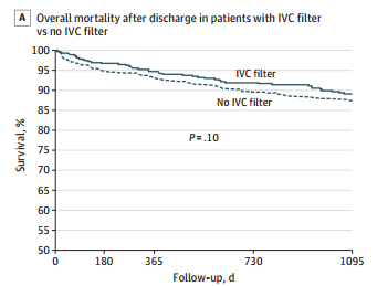
  • Over 18,000 patients were admitted during the study period, resulting in 451 with an IVC filter inserted and 1343 matched controls
  • The patients were followed for an average of 4 years after hospitalization
  • Mortality was identical between patients with filters vs the matched controls

dvt-study

  • There was still no difference in mortality, even if the patients with the filter had DVT or PE present when it was inserted
  • Only 8% ever had their “removable” filter removed (!)

Bottom line: Hopefully, it’s becoming obvious to all that the era of the IVC filter has come and gone. There are many studies that show the downside of placement. And there are several (including this one) that show how forgetful we are about taking them out when no longer needed. And, of course, they are expensive. But the final straw is that they do not seem to protect our patients like we thought (hoped?) they would. It’s time to reconsider those DVT/PE protocols and think really hard about whether we should be inserting IVC filters in trauma patients at all.

Tomorrow: a look at trends in filter insertion and retrieval.

Related post:

Reference: Association Between Inferior Vena Cava Filter Insertion
in Trauma Patients and In-Hospital and Overall Mortality. JAMA Surg, online ahead of print, September 28, 2016.

REBOA At An Academic Trauma Center

Resuscitative endovascular balloon occlusion of the aorta (REBOA) is the big thing these days. I’ve written about this topic in the past, and a number of centers continue to refine our understanding of this new(er) tool.  A recent paper from the University of Florida – Gainesville outlines their experience in implementing this procedure at an academic Level I trauma center.

This trauma program is staffed by a group of surgeons who have considerable experience in guidewire-based skills, fellowship or military exposure, and/or completion of a vascular fellowship. One surgeon attended a trauma endovascular skills course (6 hrs).  An internal education program with a 1.5 hour slide presentation and some hands-on simulation training was developed. All surgeons and residents completed this program.

A retrospective review of their experience from June 2015 to March 2017 was carried out on unstable trauma patients due to hemorrhage. All cases were performed in a hybrid OR with imaging capabilities. A 12Fr REBOA catheter was initially used, but was changed to 7Fr once that catheter became commercially available.

Here are the factoids:

  • 16 patients underwent REBOA in this 22 month period; mean SBP was 97 torr and mean ISS was 39
  • Hemodynamic status improved in 10 of 16 patients to a mean SBP 132
  • 14 survived the initial operative procedure, but only 6 survived to hospital day 30. It appears that all of these patients were neurologically normal (GCS 15+0).
  • 1 survivor developed a common femoral artery pseudoaneurysm
  • The authors made the interesting comment that they also performed 8 ED thoracotomies (EDT) during this period and that there were no survivors
  • The authors concluded that the procedure was beneficial, that extensive training was not needed, and that it should be available trauma centers

Bottom line: But not so fast! This was a very select academic Level I center. The surgeons had extensive wire skills and vascular experience. All procedures were performed in a hybrid room, which is a very controlled OR setting. And they only performed REBOA every 6 weeks or so. 

REBOA is still an advanced procedure, and the average trauma surgeon would probably benefit from some more intensive training to ensure adequate initial skills. But if the surgeon can’t then maintain their skills via somewhat regular practice, errors may creep in. In a group of 6-8 surgeons, each may only get to perform the procedure once a year! Add in some interested emergency physicians, and no one can keep in practice.

The bit about ED thoracotomy is a bit of a red herring. Typically, this procedure is performed once the patient has lost their vital signs. Comparing mortality from REBOA with EDT here is not valid, because it appears that most of the REBOA patients in this study still had vital signs when it was inserted. It would be interesting if the authors shared the outcomes in the REBOA patients who had the device inserted after arrest to level the playing field with EDT.

So what to do? Be cautious and thorough if you are planning to try out REBOA at your center. Do the math. On how many patients per year can I expect to perform this? How many physicians want credentialing to do it? How many procedures can the typical physician expect per year? What is the baseline level of physician training and what additional training is needed? Will I report my experience to a national registry or write it up for sharing?

These are important questions! Everyone wants to play with the newest shiny toy in the toybox. But make sure that when you do play with it, you are able to provide the maximum benefit to your patients with the least amount of harm!