Category Archives: Pop quiz

What Would You Do? Teensy Weensy Stab To The Abdomen – Part 3

In my last post, I described the plight of a young man who had sustained a stab to the abdomen. It appeared that a very tiny bit of omentum was hanging out of the wound. What to do?

I listed three options:

  • Local wound exploration
  • CT scan of the abdomen
  • Proceed to the operating room

So let’s work through these. First, local wound exploration (LWE).

LWE is a diagnostic procedure to determine if a sharp wound has actually or potentially penetrated a vital area. It is usually performed in the neck to determine if the platysma has been violated, or in the abdomen top check for peritoneal violation. In this case, you would use it if you just couldn’t believe that the bit of odd fat was actually omentum, or if you were unsure what you were looking at. You could also grab it (gently) and give it a little tug. If more comes out, you’ve made your diagnosis. Fortunately, this is rarely necessary because omentum has a very distinctive appearance. You know it when you see it.

What about probing the wound? One of my mentors, John Weigelt, used to ask, “Michael, does your finger / q-tip / instrument have an eyeball on the end of it?” His point was that probing is like so many other medical tests: diagnostic if positive, but unsettling if it’s not. What happens if the wound does penetrate, but you can’t find the path that the knife/bullet took? You can only call that indeterminate. I suppose you could take an approach that includes probing first, then proceeding to full LWE if that is negative.

I’ll describe the proper technique for local wound exploration in a later post.

And what about CT scan? This is another unsatisfying test, because it is very likely to be negative with small wounds. The fascial defect in this case will be very small, and can easily be missed on the scan. Not recommended.

Given all this discussion, my vote is to proceed to the operating room. I know this is omentum, and I know that there is a good likelihood that there will be an injury that needs repair. So let’s go get it done.  But what procedure should I do, and how should I do it? That’s the subject for my next post.

As always, please leave comments below or tweet them out!


What Would You Do? Teensy Weensy Stab To The Abdomen – Part 2

Yesterday, I presented the case of a young man with a teensy weensy little stab to his abdomen, just above the umbilicus. There was a tiny bit of oddly colored fat that was visible in the wound. So now what should we do?

The first thing is to figure out what that bit of fat is. It doesn’t have the normal large pebbling and color of subcutaneous fat. Therefore, it must be a small piece of omentum protruding from the wound.

And what is the significance of that? This question has been addressed by papers with low numbers of subjects since the 1980s. It really depends on what country you are located in. Do you have readily available OR resources? Are there pressures to minimize hospital stays (US)?

One of the earliest papers originated from Parkland Hospital in Dallas TX. They reviewed 115 cases of omental evisceration over a 4 year period, and found that “serious” abdominal injuries were found in 75% of them. All went to laparotomy, and injuries to not one, but two organs were noted in about half of the positive cases. There was a 7% complication rate with negative laparotomy,

Contrast this with a study from Kingston, Jamaica where 66 patients with abdominal stabs and omental evisceration were treated. Of these, 14 were treated with observation because they had a normal abdominal exam. All were treated successfully without operation. But note the ratio here: 14/66 = 21%, which is the same as the negative laparotomy in the Parkland study (25%). So this study implies that if the patient can be watched and does not develop symptoms, the negative lap may be avoided.

Unfortunately, in many countries there are pressures to get people out of the hospital as soon as possible. Since small bowel content is relatively benign (at first), patients may not become symptomatic for several days. It would probably be difficult to convince your hospital to keep patients laying around for serial exams for days on end. Not to mention the logistical problems of doing good serial exams.

So most trauma professionals will be compelled to do something. And what should we do? Here are some possibilities. Pick your poison, and I’ll give you my choice tomorrow.

  • Local wound exploration
  • CT scan of the abdomen
  • Proceed to the operating room

As before, leave a comment to let me know what you would do. Or tweet it out!

References:

  1. Significance of omental evisceration in abdominal stab wounds. Am J Surg 152(6):670-673, 1986.
  2. Non-operative management of stab wounds to the abdomen with omental evisceration. J Royal Col Surg Edin 41(4):239-240, 1996.

What Wound You Do? A Teensy Weensy Stab To The Abdomen

Here’s a case to test your mettle! A young male walks into the triage desk in your ED with a teensy weensy little puncture just above his umbilicus. Your triage nurse, who is very astute, recognizes that this meets your trauma activation criteria and pushes the button. The gentleman is escorted to your trauma bay and the team quickly assembles to evaluate him.

Vital signs are stable, and no other wounds are found. There is a very small 1cm stab located about 2cm above the umbilicus, perfectly in the midline. The abdomen is soft and nontender, and the patient wants to know why everyone is making such a big deal about this.

Upon close inspection of the wound, there is a very small piece of bright yellow fat protruding 2mm from the wound. It somehow doesn’t look like the subcutaneous fat around it.

Here are the questions that I’ll be addressing over the next several posts:

  • What do you think of the appearance of the patient and his wound?
  • Where should we go next?
  • What are our diagnosis and management options?

In my next post, we’ll discuss how we diagnose this patient and whether there is a real problem here.

What do you think is going on? What is it? What do we do next? Leave a comment here, or tweet out your answers before tomorrow!

Pop Quiz: What’s The Diagnosis? The Answer

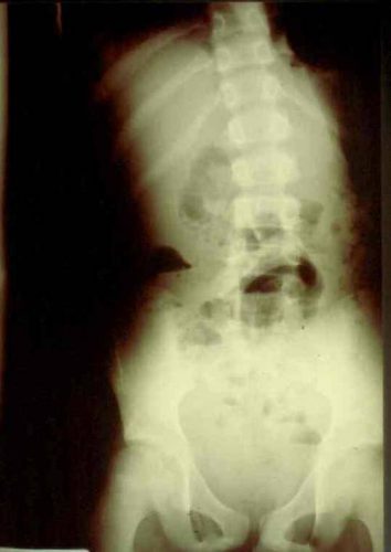
Okay, time for the answer. This 12 year old crashed his moped, taking handlebar to the mid-epigastrium. Over the next 3 days, he felt progressively worse and finally couldn’t keep food down.

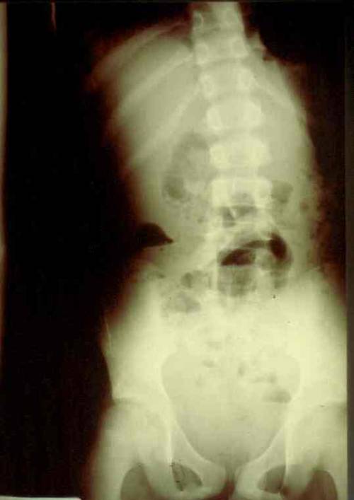
Mom brought him to the ED. The child appeared ill, and had a WBC count of 18,000. The abdomen was firm, with involuntary guarding throughout and a hint of peritonitis. The diagnosis was made on the single abdominal xray shown yesterday. Here is a close-up of the good stuff?

Emergency docs, your differential diagnosis list with this history is a pancreatic vs a duodenal injury based on the mechanism.

Classic findings for duodenal injury:

  • Scoliosis with the concavity to the right. This is caused by psoas muscle irritation and spasm from retroperitoneal soiling by the duodenal leak.
  • Loss of the psoas shadow on the right. Hard to see on this xray, but the left psoas shadow is visible, the right is not. This is due to fluid and inflammation along this plane.
  • Air in the retroperitoneum. In this closeup, you can actually see tiny bubbles of leaked air outlining the right kidney. There are also bubbles along the duodenum and a few along the right psoas.

We fluid resuscitated first (important! dehydration is common and can lead to hemodynamic issues upon induction of anesthesia) and performed a laparotomy. There was a  blowout in the classic position, at the junction of 1st and 2nd portions of the duodenum. The hole was repaired in layers and a pyloric exclusion was performed, with 2 closed drains placed in the area of the leak.

The child did well, and went home after 5 days with the drains out. Feel free to common or leave questions!