Category Archives: Pop quiz

Pop Quiz: What’s The Diagnosis? The Answer

Okay, time for the answer. This 12 year old crashed his moped, taking handlebar to the mid-epigastrium. Over the next 3 days, he felt progressively worse and finally couldn’t keep food down.

Mom brought him to the ED. The child appeared ill, and had a WBC count of 18,000. The abdomen was firm, with involuntary guarding throughout and a hint of peritonitis. The diagnosis was made on the single abdominal xray shown yesterday. Here is a close-up of the good stuff?

Emergency docs, your differential diagnosis list with this history is a pancreatic vs a duodenal injury based on the mechanism.

Classic findings for duodenal injury:

  • Scoliosis with the concavity to the right. This is caused by psoas muscle irritation and spasm from retroperitoneal soiling by the duodenal leak.
  • Loss of the psoas shadow on the right. Hard to see on this xray, but the left psoas shadow is visible, the right is not. This is due to fluid and inflammation along this plane.
  • Air in the retroperitoneum. In this closeup, you can actually see tiny bubbles of leaked air outlining the right kidney. There are also bubbles along the duodenum and a few along the right psoas.

We fluid resuscitated first (important! dehydration is common and can lead to hemodynamic issues upon induction of anesthesia) and performed a laparotomy. There was a  blowout in the classic position, at the junction of 1st and 2nd portions of the duodenum. The hole was repaired in layers and a pyloric exclusion was performed, with 2 closed drains placed in the area of the leak.

The child did well, and went home after 5 days with the drains out. Feel free to common or leave questions!

Knife To The Back – The Conclusion!

To summarize: stab to the back, prone position, stable vitals, awake and alert and breathing easily. The patient had a chest xray which showed some likely hemothorax. He was sent to CT (prone) and the image obtained looked like this:

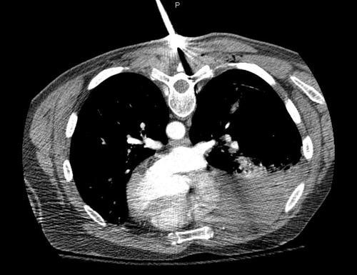
They key points to note are:

  • The injury is completely above the diaphragm. No need to worry about an intra-abdominal problem.
  • The amount of hemothorax is moderate. It is not enough to mandate a thoracotomy. At least for now.
  • There is a significant pneumothorax. You can’t see it due to the windows used, but the lung has separated from the chest wall by about 3cm.
  • The track of the knife was directed laterally.
  • No significant vascular structures were involved, and there is no contrast extravasation.

Final management: The patient was returned to the ED, and the knife was deftly removed and processed properly as evidence. The patient was then turned supine and a 40 Fr chest tube was inserted using procedural sedation. About 400 cc of blood was drained and reinfused. A repeat chest xray was obtained, which showed some residual hemothorax and near resolution of the pneumothorax. He was then admitted for frequent vital signs and drainage measurements for two shifts. Afterwards, he was placed in our chest tube management protocol. The tube was removed and he was discharged two days later. There were no complications.