Category Archives: Guidelines

A New Proposed Practice Guideline For Cervical Spine Clearance

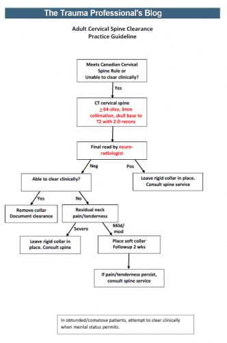
In my last post, I reviewed a very recent prospective study on using CT scan alone for  cervical spine clearance in intoxicated patients. I believe that this is the final piece in the spine clearance puzzle to allow us to perform this task intelligently.

We’ve been accumulating more and more data that supports the use of CT scan in patients who fail clinical clearance. This failure can be due to the patient being obtunded or intoxicated, bearing a “distracting” injury, or being just plain uncooperative. Because of this, and our fear of missing a potentially devastating injury (typically because of rare anecdotal cases or urban legends), we have resorted to a significant degree of overkill. This has included, over the years, prolonged immobilization in a rigid collar, flexion/extension imaging (plain x-ray or fluoro), and MRI.

I’ve synthesized the available literature, and have drafted a simple, one sheet practice guideline for discussion. In order to use it, you must have the following:

  • A decent CT scanner – minimum 64 slice
  • A well-defined scan setup protocol – 3mm collimation, skull base to T2, 2-D reconstruction in sagittal and coronal planes (get a copy of our protocol below)
  • A skilled radiologist – neuroradiologist required

An image of the protocol can be found at the bottom of this post. I’m interested in your comments, and your comfort or discomfort with adopting something like this. Please leave comments here or on twitter.

Links: 

Reference: Cervical spine evaluation and clearance in the intoxicated patient: A prospective Western Trauma Association Multi-Institutional Trial and Survey. J Trauma 83(6):1032-1040, 2017.

Print Friendly, PDF & Email

It’s Time To Simplify Cervical Spine Clearance!

Cervical spine clearance is another one of those tasks that everyone seems to do their own way. Most trauma centers have an algorithm for clearance, or even two, like my center. But anytime different clinicians or centers do the same thing in different ways, it means we don’t really know what we’re doing. 

It basically means that the hard data is not there to dictate what we truly should do. So there are two alternatives:

  1. Wait for good data to become available. Unfortunately, this can take forever.
  2. Extrapolate from any existing data, and fill in the gaps with our clinical experience to come up with something that works and causes no harm.

The protocols in use at Regions Hospital are based on #2, and have been in place for over a decade. But now, we have a good example of #1 to work with.

Fortunately for us, cervical spine clearance has been evolving for decades. And as technology has improved, so has our ability to miss fewer and fewer “significant” injuries. A multi-center trial published this month provides one of the final puzzle pieces to help us settle upon a uniform cervical spine clearance guideline. It was a prospective look at intoxicated patients after blunt trauma, who can’t always participate in the process of clinical cervical spine clearance.

This three year study took place at 17 centers and specifically looked at the combination of clinical and radiographic clearance in alcohol and drug intoxicated patients. Over 10,000 patients participated in the study. There are some limitations, of course, when so many centers participate. But the pros massively outweigh the cons.

Here are the factoids:

  • The overall incidence of cervical spine injury was 10.6% (!)
  • 30% of patients were intoxicated (19% etoh, 6% drugs, 5% both (also !)
  • Intoxicated patients had a significantly lower incidence of cervical injury (8% vs 12%). (Don’t get any ideas about the old adage about being relaxed when they crash. This probably represents lower speeds involved.)
  • For intoxicated patients, sensitivity of CT scan was 94%, specificity was 99.5%, and the negative predictive value (NPV) was 99.5%
  • The NPV for clinically significant injuries in intoxicated patients was 99.9%, and no unstable injuries were missed by CT  (100% NPV) (!!)
  • When CT was negative, being intoxicated led to longer time in a collar (8 hrs vs 2 hrs)

Bottom line: Fear of clearing the cervical spine without a clinical exam, or in obtunded or intoxicated patients, is primarily due to old anecdotal reports. And much of it is not first-hand experience, but rumors of others’. What is finally becoming clear is that it is okay to clear based upon radiographic findings alone. 

Tomorrow, I’ll provide my version of a new, unified clearance protocol based on this work.

Reference: Cervical spine evaluation and clearance in the intoxicated patient: A prospective Western Trauma Association Multi-Institutional Trial and Survey. J Trauma 83(6):1032-1040, 2017.

Print Friendly, PDF & Email

Retained Hemothorax Part 4: The Practice Guideline

Over the last three days, I reviewed some data on lytics at the request of some of my readers. Then I looked at a paper describing one institution’s experience dealing with retained hemothorax, including the use of VATS. But there really isn’t much out there on how to roll all this together.

Until now. The trauma group at Vanderbilt has a paper in press describing their experience with a home-grown practice guideline for managing retained hemothorax.  Here’s what it looks like:

I know it’s small, so just click it to download a pdf copy. I’ve simplified the flow a little as well.

All stable patients with hemothorax admitted to the trauma service were included over a 2.5 year period. The practice guideline was implemented midway through this study period. Before implementation, patients were treated at the discretion of the surgeon. Afterwards, the practice guideline was followed.

Here are the factoids:

  • There were an equal number of patients pre- and post-guideline implementation (326 vs 316)
  • An equal proportion of each group required an initial intervention, generally a chest tube (69% vs 65%)
  • The number of patients requiring an additional intervention (chest tube, VATS, lytics, etc) decreased significantly from 15% to 9%
  • Empyema rate was unchanged at 2.5%
  • Use of VATS decreased significantly from 8% to 3%
  • Use of catheter guided drainage increased significantly from 0.6% to 3%
  • Hospital length of stay was the same, ranging from 4 to 11 days (much shorter than the lytics studies!)

Bottom line: This is how design of practice guidelines is supposed to work. Identify a problem, typically a clinical issue with a large amount of provider care variability. Look at the literature. In general, find it of little help. Design a practical guideline that covers the major issues. Implement, monitor, and analyze. Tweak as necessary based on lessons learned. If you wait for the definitive study to guide you, you’ll be waiting for a long time.

This study did not significantly change outcomes like hospital stay or complications. But it did decrease the number of more invasive procedures and decreased variability of care, with the attendant benefits from both of these. It also dictates more selective (and intelligent) use of additional tubes, catheters, and lytics. 

I like this so much that I plan to adopt it at my center!

Download the practice guideline here.

Related posts:

Posts in this series:

Reference: Use of an evidence-based algorithm for patients with traumatic hemothorax reduces need for additional interventions. J Trauma, in press, December 14, 2016.

Print Friendly, PDF & Email

ED Thoracotomy: Practice Management Guideline

I still encounter a quite a bit of confusion about ED thoracotomy: when to do it, when to stop. A nice set of guidelines was developed by the Subcommittee on Outcomes of the American College of Surgeons Committee on Trauma about 10 years ago. And really, things haven’t changed very much since then.

In general, the literature on this topic has been all over the place. Most studies are retrospective with very little statistical validation. But there’s a lot of it out there. The subcommittee used fairly standard methods to evaluate the literature and come up with some recommendations.

The overall survival rate for all comers was 8% (11% for penetrating, 1.6% for blunt). About 15% of survivors (no breakdown of blunt vs penetrating) suffered from neurologic impairment. Penetrating cardiac injury had the best survival (31%). In the 4 studies on children, the survival numbers paralleled the adults.

Because of the relatively weak quality of the data, only level II recommendations were given. They were:

  • Don’t consider ED thoracotomy in blunt trauma patients unless the patient arrests in front of you. Otherwise, meaningful survival is almost nonexistent.
  • Consider ED thoracotomy for penetrating chest injury if there were witnessed signs of life and a short transport time (5-10 minutes max, in my opinion). Look for pupillary response, spontaneous respirations, palpable pulse, or a narrow complex cardiac rhythm.
  • ED thoracotomy for penetrating abdominal injuries has a low survival rate, but can be considered if the injury may involve the chest.
  • Consider thoracotomy for suspected abdominal vascular injury, but survival is also very low.
  • The guidelines above apply equally to children.

Practical tips: ED thoracotomy is a seldom used and dangerous procedure. There are many opportunities for injury to trauma professionals, so be selective and take precautions. Assign someone to run the chemical code while the surgeons open the chest. Watch out for broken ribs and scalpels gone wild!

I’ll post pictures and specific pointers over the next three days.

Related posts:

Reference: Practice management guidelines for emergency department thoracotomy. JACS 193(3):303-309, 2001.

Print Friendly, PDF & Email