Category Archives: Guidelines

Massive Transfusion: What Ratios Are People Using?

This is the first of a two-part series on massive transfusion protocol (MTP) ratios. Today, I’ll write about what ratios trauma centers around the country are using. Tomorrow I’ll review the literature we have to date on what the correct ratio should be. Are we all doing the right thing or not?

Back in the old days (which I remember fondly), we didn’t pay too much attention to the ratio of blood to plasma. We gave a bunch of bags of red cells, then at some point we remembered that we should give some plasma. And platelets? We were lucky to give any! And to top it all off, we gave LOTS of crystalloid. Turns out this was not exactly the best practice.

But things have changed. Some good research has shown us that a nice mix of blood component products is good and too much crystalloid is bad. But what exactly is the ideal mix of blood products? And what is everybody else doing? I’ll try to answer these questions in this series.

So first, what are all the other trauma centers doing? An interesting medley of anesthesia and pathology groups from the University of Chicago, a Dallas-based anesthesia group, and a blood center in my home base of St. Paul, conducted a survey of academic medical centers in 2016. They wanted to find out how many actually had a MTP and to scrutinize the details.

They constructed a SurveyMonkey survey and sent it to hospitals with accredited pathology residencies across the US. There were 32 questions in the survey, which asked for a lot of detail. As you can probably personally attest, the longer and more complicated the survey, the less likely you are to respond. That certainly happened here. Of 107 surveys sent out, it took a lot of nagging (initial email plus two nags) to get a total of 56 back.

Here are the factoids:

  • Most were larger hospitals, with 74% having 500 or more beds
  • All had massive transfusion protocols
  • Trauma center level: Level I (77%), Level II (4%), Level III (4%), Level IV (2%), no level (14%)
  • Nearly all (98%) used a fixed ratio MTP; very few used any lab-directed (e.g. TEG/ROTEM) resuscitation
  • Target RBC:plasma ratio: 1:1 (70%), 1.5:1 (9%), 2:1 (9%), other (9%)
  • Only 58% had the same RBC:plasma ratio in each MTP cooler
  • More than 86% had thawed plasma available (remember, these were generally large academic centers)
  • Half stored uncrossmatched type O PRBCs outside the blood bank, usually in the ED; only 1 stored thawed plasma in the ED
  • A total of 41% had more than one MTP (trauma, OB, GI, etc.)
  • 84% had some type of formal review process once the MTP was complete
  • About 68% had modified their MTP since the original implementation. Some increased or decreased ratios, expanded MTP to non-trauma services, decreased the number of units in each pack, changed to group A plasma from AB, or switched from ratio to TEG/ROTEM or back.

Bottom line: This is an intriguing snapshot of MTP practices around the country that is about four years old. Also remember, this is a somewhat skewed dataset. The survey was directed toward hospitals with academic pathology programs, not trauma centers. However, there is enough overlap that the results are probably generalizable. 

Most centers are (were) using MTP packs containing six units of PRBCs, and were attempting to achieve a fixed 1:1 ratio. Half of hospitals had the same number of units in each cooler, half varied them by cooler number. Nearly half had multiple flavors of MTP for different specialties. Very few used TEG/ROTEM during the initial phased of MTP. Most modified their MTP over time.

I’ve written quite a lot on most of these issues. See the links to my “MTP Week” series from earlier this year, below.

Tomorrow, I’ll review what we know and don’t know about the proper ratios to use in your MTP.

Reference: Massive Transfusion Protocols: A Survey of Academic
Medical Centers in the United States. Anesth & Analg 124(1):277-281, 2017.

MTP week series:

Best Practice: Use of CT Scan In Trauma Activations – Part 2

In my last post, I described how the unscheduled and random use of CT scan in trauma activations can interfere with normal radiology department workflow, creating access problems for other emergency and elective patients. Today, I’ll detail a project implemented at my hospital to analyze the magnitude of this problem and try to resolve it.

We started with a detailed analysis of how the scanner was being used for trauma activation patients. Regions Hospital has a single-tier trauma activation system, with no mechanism of injury criteria other than penetrating injury to the head, neck, and torso. There are usually about 850 activations per year, and traditionally the CT scanner has been “locked down” when the activation is announced. The CT techs would complete the current study on the table, then hold the scanner open until called or released by the trauma team.

Since we are a predominantly blunt trauma institution, we scan most stable patients. Our average time in the trauma bay is a bit less than 20 minutes. Add this time to the trauma activation prenotification time of up to 10 minutes, and the scanner has the potential to sit idle for up to half an hour. And in some cases when scan is not needed (minor injuries, rapid transport to OR) the techs were not notified and were not aware they could continue scanning their scheduled cases.

A multidisciplinary group was created and started with direct observation of the trauma activation process and a review of chart documentation and radiology logs. On average it was calculated that the scanner was held idle for an average of 17.9 minutes too long. This is more than enough time to complete one, or even two studies!

A new process was implemented that required the trauma team leader to call out to the ED clerk placing orders for the resuscitation 5 minutes before the patient would be ready for scan. I still remember the first time this happened to me. I was so used to just packing up and heading to scan, I got a little irritated when told that I hadn’t made the 5-minute call. But it’s a good feedback loop, and I never forgot again!

We studied our workflow and results over a 9-week period. And here are the factoids:

  • The average CT idle time for trauma activations before the project was 17.9 minutes
  • This decreased to an average idle time of 6.4 minutes during the pilot project
  • Total idle time for all activations was 8.3 hours, but would have been 36 hours under the old system
  • A total of 28.6 hours were freed up, which allowed an additional 114 patients to be scanned while waiting for the trauma activation patients

This was deemed a success, and the 5-minute rule is now part of the routine flow of our trauma activations. We rarely ever have to wait for CT, and if we do it’s usually due to the team leader not thinking ahead.

Bottom line: This illustrates the processes that should be used when a quality problem surfaces in your program:

  • Recognize that there is a problem
  • Convene a small group of experts to consider the nuances
  • Generate objective data that describes the problem in detail
  • Put on your thinking caps to come up with creative solutions
  • Test the solutions until you find one that shows the desired improvement
  • Be prepared to modify your new systems over time to ensure they continue to meet your needs

Best Practice: Use of CT Scan In Trauma Activations – Part 1

Computed tomography is an essential part of the diagnostic workup for many trauma patients. However, it’s a limited resource in most hospitals. Only so many scanners are affordable and available.  Typically, trauma centers have a scanner located in or very near the trauma bay, which makes physical access easy. Others may be located farther away, which can pose logistical and safety issues for critically injured patients.

Even if the CT is close to the ED, availability can be an issue. This availability applies not only to trauma scans, but to others as well. There is an expectation that CT be immediately available when needed for trauma activation patients. However, chances are that the same scanner is also used for high priority scans for services other than trauma, such as stroke evaluation.

Who gets the scanner first? Obviously, many trauma patients need rapid diagnosis for treatment of their serious injuries. But a fresh stroke patient also has a neurologic recovery countdown clock running if they might be eligible for lytic administration.

And don’t forget that trauma and stroke aren’t the only services vying for that scanner. The hospital undoubtedly has a stream of elective scans queued up for other in-house patients. Every urgent or emergent scan needed for trauma sets the elective schedule back another 30 minutes or more.

How does your trauma center manage CT scan usage for trauma? The vast majority essentially lock it down at some fixed point. This is typically either upon trauma activation, or at patient arrival. The former is very common, but also very wasteful because there can be a significant wait for the patient to actually arrive. Then add on the time it takes to complete the trauma bay evaluation. Up to an hour may pass, with no throughput in the CT scanner. This can be a major work flow headache for your radiology department.

Is there another way? My center was one of those that stopped the scanner after the current patient was finished at the time the trauma activation was called. We have two scanners just 30 feet from the trauma bays, so one could continue working while the other was held. However, this cut their throughput by 50% for roughly half an hour. We recognized that this was a creating a problem for the whole hospital, so we worked with the radiology department to come up with a better way.

Tomorrow I’ll detail the new system we implemented, and provide data showing the real impact of this new system on CT scan productivity.

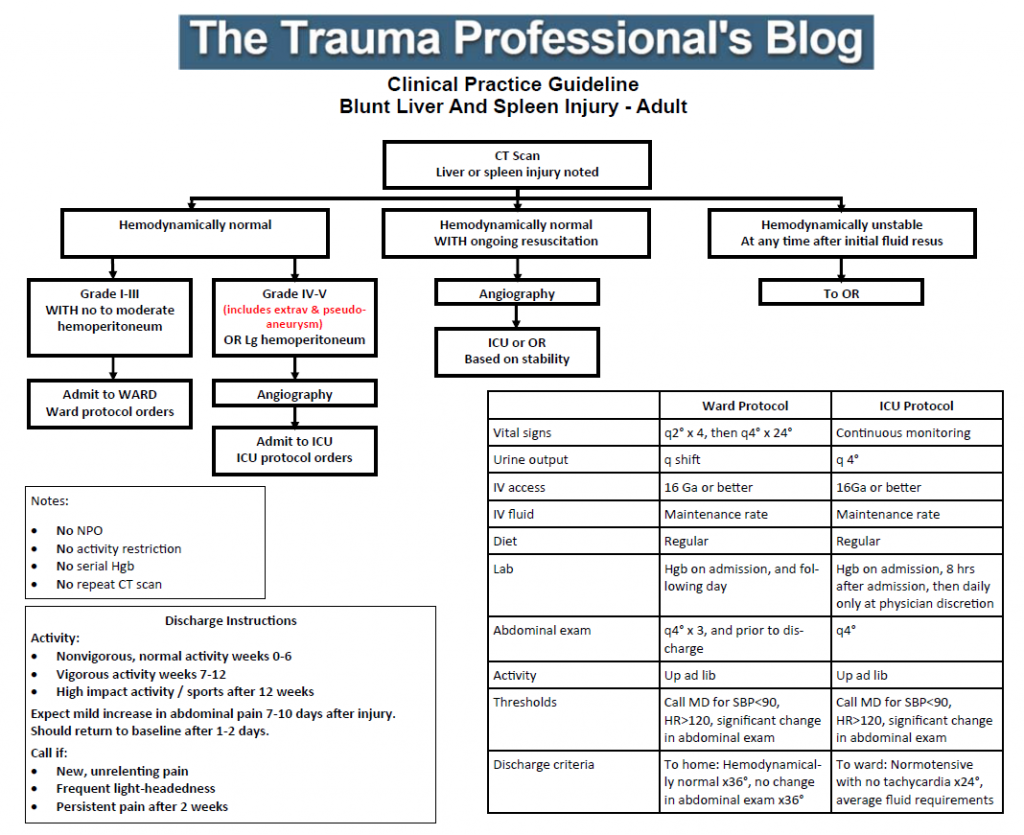
Solid Organ Injury Practice Guideline Updated

Regions Hospital developed a clinical practice guideline for solid organ management in 2002-2003. It has been revised a few times over the years, as any good guideline should with the availability of new data.

I’ve just put the finishing touches on the latest revision as a result of the updated organ scaling rules published by the American Association for the Surgery of Trauma. I reviewed the new scales for both liver and spleen earlier this year (links below). In the previous iteration of the scaling system, the importance of contrast pooling (pseudoaneurysm) or extravasation beyond the organ was not well defined. 

The new guideline explicitly includes these injuries in the high grade group, which for us is grade IV or V. Technically, pseudoaneurysm of the liver is only grade III, but in my opinion demands angiographic investigation and embolism. Thus the inclusion in the high grade / angiography arm of our guideline.

For those of you who have not seen this guideline before, there are several important directives that are listed on the left side of the page:

  • Patients are NOT made NPO
  • They do NOT have activity restrictions (such as bed rest)
  • Serial hemoglins are NOT drawn
  • An abdominal CT scan is NOT repeated

These changes were made in 2015 based on our clinical experience that properly selected patients almost never failAnd they still don’t, so why starve, restrain, poke, and re-irradiate them?

Additionally, we included explicit impact activity restrictions for post-discharge so that patients would get the same message from all members of our team.

Click the image below to download the guideline and have a look. I’m interested in your comments!

Related posts:

Guidelines for Consultants to the Trauma Service

This post is a favorite, and I’m publishing it again since I just finished my “When To Call” series.

Trauma surgeons often rely on consultants to assist in the care of their patients. Orthopedic surgeons and neurosurgeons are some of the more frequent consultants, but a variety of other surgical and medical specialists may be needed. I have found that providing a set of guidelines to consultants helps to ensure quality care and provide good communication between caregivers and patients / families.

We have disseminated a set of guidelines to our colleagues, and I wanted to touch on some of the main points. You can download the full document using the link at the bottom of this post.

In order to deliver the highest quality and most cost-effective care, we request that services we consult do the following:

  • Please introduce yourself to our patient and their family, and explain why you are seeing them.
  • Although you may discuss your findings with the patient, please discuss all recommendations with a member of the trauma service first. This avoids patient confusion if the trauma team chooses not to implement any recommendations due to other patient factors you may not be aware of.
  • Document your consultation results in writing (paper or EMR) in a timely manner.
  • If additional tests, imaging or medications are recommended, discuss with the trauma service first. We will write the orders or clear you to do so if appropriate, and will discuss the plan with the patient.
  • We round at specific times every day and welcome your attendance and input.
  • Please communicate any post-discharge instructions to us or enter in the medical record so we can expedite the discharge process and ensure all followup visits are scheduled.

Bottom line: A uniform “code of behavior” is important! Ensuring good patient communication is paramount. They need to hear the same plans from all of their caregivers or else they will lose faith in us. One of the most important lessons I have learned over the years is that you do not need to implement every recommendation that a consultant makes. They may not be aware of the most current trauma literature, and they will not be familiar with how their recommendations may impact other injuries.

Click here to download the full copy of the Regions Hospital Trauma Services consultant guidelines.