Category Archives: General

Dysphagia and Cervical Spine Injury

Cervical spine injury presents a host of problems, but one of the least appreciated ones is dysphagia. Many clinicians don’t even think of it, but it is a relatively common problem, especially in the elderly. Swallowing difficulties may arise for several reasons:

  • Prevertebral soft tissue swelling may occur with high cervical spine injuries, leading to changes in the architecture of the posterior pharynx
  • Rigid cervical collars, such as the Miami J and Aspen, and halo vests all force the neck into a neutral position. Elderly patients may have a natural kyphosis, and this change in positioning may interfere with swallowing. Try extending your neck by about 30 degrees and see how much more difficult it is to swallow.
  • Patients with cervical fractures more commonly need a tracheostomy for ventilatory support and/or have a head injury, and these are well known culprits in dysphagia

A study in the Jan 2011 Journal of Trauma outlines the dysphagia problem seen with placement of a halo vest. They studied a series of 79 of their patients who were treated with a halo. A full 66% had problems with their swallowing evaluation. This problem was associated with a significantly longer ICU stay and a somewhat longer overall hospital stay.

Bottom line: Suspect dysphagia in all patients with cervical fractures, especially the elderly. Carry out a formal swallowing evaluation, and adjust the collar or halo if appropriate. 

Reference: Swallowing dysfunction in trauma patients with cervical spine fractures treated with halo-vest fixation. J Trauma 70(1):46-50, 2011.

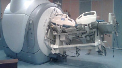
MRI And External Fixators

MRI is an indispensable tool for evaluation of spine and soft tissue trauma. However, a great deal of effort was be made to ensure that any patient scheduled for this test is “MRI compatible.” The fear is that any retained metallic fragments may move or heat up once the magnets are activated. 

But what about trauma patients with external fixators? That is one big hunk of metal that is inserted deep into your patient. There are three major concerns:

  • Is the material ferromagnetic? If so, it will move when the magnets are activated and may cause internal injury. These days, there are many fixator sets that are not ferromagnetic, avoiding this problem.
  • Can currents be induced in the material, causing heating? This is not much of a problem for small, isolated objects. However, external fixators are configured in such a way that loops are created. The fluctuating magnetic fields can induce currents that in turn will heat the surrounding tissue. And thinner materials (narrow pins) result in more current and more heating.
  • Will the metal degrade image quality?

The biggest challenge is that there is no standard ex-fix configuration. Our orthopaedic colleagues get to unleash their creativity trying to devise the appropriate architecture to hold bones together so they can heal properly. This makes it difficult to develop standardized guidelines regarding what can and can’t go into the scanner.

However, there is a growing body of literature showing that the heating effects are relatively small, and get smaller as the distance from the magnet increases. And non-ferromagnetic materials move very little, if at all, and do not interfere with the image. So as long as nonferromagnetic materials are used, the patients are probably safe as long as basic principles are adhered to:

  • Other diagnostic options should be exhausted prior to using MRI.
  • Informed consent must be obtained, explaining that the potential risks are not completely understood.
  • The fixator must be tested with a handheld magnet so that all ferromagnetic components can be identified and removed.
  • All traction bows must be removed.
  • Ice bags are placed at all skin-pin interfaces.
  • The external fixator must remain at least 7cm outside the bore at all times.

Bottom line: MRI of patients with external fixators can be safely accomplished. Consult your radiologists and physicists to develop a policy that is specific to the scanners used at your hospital. 

Related posts:

Obit: Erwin Thal MD – 1936-2014

Another giant has passed from the trauma world. Erwin Thal died Saturday morning in Dallas, his home city. I have known this brilliant and charming surgeon for many years, and always enjoyed his company and his fine wit. It saddens me greatly that we will no longer feel his direct influence on our field.

image

Dr. Thal attended my medical school alma mater, The Ohio State University. He went on to complete his internship and residency at Parkland Memorial Hospital in 1969. He then joined the faculty there and has been an integral part of that program for the past 45 years. He co-authored numerous papers and book chapters, and was a major presence in the education of trauma and surgical trainees.

His presence will be missed but never forgotten.

Trauma Coverage By Locum Tenens Surgeons

Trauma call coverage is not always easy to come by, especially at lower level trauma centers and in rural areas. Many centers come to rely on locum tenens surgeons to fill gaps in their call schedules. Unfortunately, this can create some headaches.

There is currently no trauma literature on this topic. Other disciplines, most recently pediatric surgery, have some published suggestions (I hesitate to call them guidelines) on requirements and expectations based on the ACGME core competencies.

Here are some of the nuances that any trauma program needs to recognize if the use of locum tenens surgeons is being considered:

  • Board certification – This is a basic tenet of trauma center verification and should be absolutely required
  • Trauma CME – Make sure that all locums meet the CME or internal education program (IEP) requirement before they start
  • Core vs non-core surgeon – Locums are best designated a non-core surgeon so they are not required to attend multidisciplinary PI committee meetings (MDPI)
  • Dissemination of committee proceedings – Make sure that this is well-documented. Since these surgeons are not required to attend MDPI if they are non-core, they must be aware of all items discussed, particularly if it involves their care
  • Responsibility for quality issues – This is the most troubling aspect of using locums. It’s tough to hold one of these surgeons responsible for issues arising from their care if they have left and are never coming back. Make sure there is a mechanism to send feedback about their care even after they are gone for good. And document it well!

Bottom line: In my opinion, the use of locum tenens to cover trauma call gaps is a necessary evil. It should only be used until a more stable coverage pool is available. The management of quality issues in particular is much more difficult when using roving surgeons. And with the adoption of the new Resource Document on July 1, 2015 (Orange Book), it’s going to get even harder to use them. If you must, use them wisely and only briefly.

Reference: Proposed standards for use of locum tenens coverage in pediatric surgery practices. J Pediatric Surg 48:700-703, 2013 (letter).

Interesting Concept: The Abdominal Aortic & Junctional Tourniquet

Tourniquets for extremity bleeding are definitely back in vogue. Our military experience over the past 20 years has shown us what a life saver this simple tool can be. It’s now carried by many prehospital trauma professionals for use in the civilian population. But what about bleeding from the nether regions? You know what I’m talking about, the so-called junctional zones. Those are the areas that are too proximal (or too dangerous) to put on a tourniquet, like the groin, perineum, axilla, and neck.

Traditionally, junctional zone injury could only be treated in the field with direct pressure, clamps, or in some cases a balloon (think 30Fr Foley catheter inserted and blown up as large as possible, see link below). In the old days, we could try blowing up the MAST trousers to try to get a little control, but those are getting hard to find. 

An Alabama company (Compression Works) developed a very novel concept to try to help, the Abdominal Aortic and Junctional Tourniquet (AAJT). Think of it as a pelvic compression device that you purposely apply too high.

image

Note the cool warning sticker at the bottom of the device!

The developers performed a small trial on 16 volunteer soldiers after doing a preliminary test on themselves (!). The device was placed around the abdomen, above the pelvis, and inflated to a maximum of 250 torr. Here are the factoids:

  • All subjects tolerated the device, and no complications occurred
  • Flow through the common femoral artery stopped in 15 of the 16 subjects
  • The subject in whom it did not work exceeded the BMI and abdominal girth parameters of the device
  • Average pain score after application was 6-7 (i.e. hurts like hell!)

Here’s a list of the criteria that preclude use of this device:

image

Bottom line: This would seem to be a very useful device for controlling hemorrhage from pesky areas below the waist.

BUT! Realistically, it will enjoy only limited use in the civilian population for now. Take a closer look at the exclusion criteria above. Half of the population is ineligible right off the bat (women). And among civilians, more than a third are obese in the US. Toss in a smattering of the other criteria, and the unlikelihood of penetrating trauma to that area in civilians, it won’t make financial sense for your average prehospital agency to carry it. Maybe in high violence urban areas, but not anywhere else.

The company has received approval for use in pelvic and axillary hemorrhage control, so we’ll see how it works when more and larger studies are released (on more and larger people). 

Related post:

Reference: The evaluation of an abdominal aortic tourniquet for the control of pelvic and lower abdominal hemorrhage. Military Med 178(11):1196-1201, 2013.

Thanks to David Beversluis for bringing this product to my attention. I have no financial interest in Compression Works.