Category Archives: General

Trauma Activation Patients Staying Too Long In Your ED?

One of the long-held beliefs in trauma care relates to the so-called “golden hour.” Patients who receive definitive care promptly do better, we are told. In most trauma centers, the bulk of this early care takes place in the emergency department. However, for a variety of reasons, throughput in the ED can be slow. Could extended periods of time spent in the ED after patient arrival have an impact on survival?

Wake Forest looked at their experience with nearly 4,000 trauma activation patients who were not taken to the OR immediately and who stayed in the ED for up to 5 hours. They looked at the impact of ED dwell time on in-hospital mortality, length of stay and ventilator days.

Overall mortality was 7%, and the average time in the ED was 3 hours and 15 minutes. The investigators set a reasonable but arbitrary threshold of 2 hours to try to get trauma activation patients out of the ED. When they looked at their numbers, they found that mortality increased (7.8% vs 4.3%) and that hospital and ICU lengths of stay were longer in the longer ED stay group. Hospital mortality increased with each hour spent in the ED, and 8.3% of patients staying between 4 and 5 hours dying. ED length of stay was an independent predictor for mortality even after correcting for ISS, RTS and age. The most common cause of death was late complications from infection.

Why is this happening? Patients staying longer in the ED between 2 and 5 hours were more badly injured but not more physiologically abnormal. This suggests that diagnostic studies or consultations were being performed. The authors speculated that the knowledge, experience and protocols used in the inpatient trauma unit were not in place in the ED, contributing to this effect.

Bottom line: This is an interesting retrospective study. It reflects the experience of only one hospital and the results could reflect specific issues found only at Wake Forest. However, shorter ED times are generally better for other reasons as well (throughput, patient satisfaction, etc). I would encourage all trauma centers to examine the flow and delivery of care for major trauma patients in the ED and to attempt to streamline those processes so the patients can move on to the inpatient trauma areas or ICU as efficiently as possible.

Reference: Emergency department length of stay is an independent predictor of hospital mortality in trauma activation patients. J Trauma 70(6):1317-1325, 2011.

Print Friendly, PDF & Email

Best Practices For TBI Patients On Oral Anticoagulants: Part 5

Here is the fifth and final installment of my series summarizing the Austrian consensus paper on management of TBI patients with intracranial hemorrhage. The previous posts have run the gamut from diagnostic tests to detection of specific drugs to management. I’ve covered platelet inhibitors and Vitamin K antagonist reversal in previous posts, and today I’ll go over the panel’s reversal strategies for the direct oral anticoagulant drugs (DOACs).

Q1. Should idarucizumab (Praxbind) always be administered to patients with hemorrhagic TBI who are taking dabigatran (Pradaxa).

Answer: Only in cases where your laboratory is not capable of testing for thrombin time.

If thrombin time (TT) can be measured and is within the normal range, then the drug is not therapeutic and reversal should not be carried out. The consensus statement recommends giving this drug if the TT is prolonged or your lab cannot measure it. Keep in mind that there are very, very few papers on DOAC reversal in trauma patients. Most studies address the stroke population, and this may not translate well to trauma. And there are no studies yet that show that idarucizumab offers any survival benefit if given.

Q2. Should prothrombin complex concentrate (PCC) always be given to patients who are taking Factor Xa inhibitors?

Answer: Only in cases where your laboratory is not capable of testing for anti-Xa activity.

If anti-Xa activity can be measured (in a timely manner) and is low, then the drug is not therapeutic and PCC need not be given. If the level is high or your lab cannot test for it, then the group recommends administering PCC if the specific reversal agent (Andexxa) is not available.

As with dabigatran above, there is very little trauma literature to justify this recommendation. Furthermore, Andexxa is very expensive and, like idarucizumab, has not been shown to improve survival. Next week, I’ll write about why Andexxa is probably not worth the cost, in my opinion.

Q3. Should DOACs always be reversed in hemorrhagic TBI?

Answer: We don’t know.

As I just mentioned, there is little if any data showing that administration of a reversal agent is beneficial. And the decision to give it must be balanced with patient risk for thrombosis and consideration of any other agents they may be taking.  Expert opinion suggests that DOACs need not be reversed in TBI without blood on the CT scan, patients with unilateral, asymptomatic chronic subdural hematoma, and those with other wounds that do not appear to be bleeding excessively.

Hopefully, this series has helped shine some light on a confusing set of problems. Next week I’ll dig a bit deeper into the DOAC reversal agents Praxbind and Andexxa.

Reference: Diagnostic and therapeutic approach in adult patients with traumatic brain injury receiving oral anticoagulant therapy: an Austrian interdisciplinary consensus statement. Crit Care 23:62, 2019.

Print Friendly, PDF & Email

Does Chest Tube Size Matter? Part 2

In my last post, I reviewed a large prospective series comparing smaller (28-32 Fr) to larger (36-40 Fr) chest tubes for management of pneumothorax. The authors did not detect any significant difference because the study was underpowered given the incidence of the adverse events examined.

Today, I’ve chosen a more recent paper that attempts to do the same thing. Interestingly, it cites the previous paper as a good example showing no differences! This one is from an emergency medicine group in Fukui, Japan. It is a retrospective review of seven years worth of patients who had a chest tube inserted for hemothorax only.

Here are the factoids:

  • Small bore tubes were 20-22 French, and large bore tubes were 28 French (huh?)
  • The tube selection was made (once again) at the discretion of the attending physician
  • Demographics and injury data from the two groups were equal
  • A total of 124 tubes were placed in 116 patients, 68 small bore and 56 large bore
  • Empyema occurred in 1% in each group
  • Retained hemothorax occurred in 2% of small tube patients and 3% of large tube patients
  • An additional tube was placed in 2% of small tube patients and 7% of large tube patients (p = 0.41)
  • Pain was not evaluated

The authors concluded that “emergent insertion of the small-bore tubes had no difference in efficacy of drainage, complications or need for additional invasive procedures.”

Bottom line: Huh? Once again we have an inferior design (retrospective review) and huge potential for selection bias (no criteria or randomization for tube size). But in this case, the tube sizes are very similar! The difference in diameter between a 20 Fr tube and a 28 Fr one is only 2.5mm! Reason #1 for no apparent differences.

For reason #2, look at the sample size. First of all, this hospital placed only 124 tubes in 7 years. That’s a one tube every three weeks. Is there that little chest trauma, or is a chunk of data missing? This sample size is less than half of that in the previous post, so the statistical power is far weaker. Look at the stats above for additional tube placement. A 3.5x change was not even close to being statistically significant. In fact, this sample size would not show a significant difference for retained hemothorax until one group had nearly 8x the number! No wonder the authors assumed there was no difference. The study was not designed in such a way that it could ever show one!

So throw this study in the trash bin, too. I’ll continue my search for a more convincing “size matters” paper in my next post.

And if you think you’ve got one, send it my way so I can have a look!

Reference: Small tube thoracostomy (20-22 Fr) in emergent management of chest trauma. Injury 48(9):1884-1887, 2017.

Print Friendly, PDF & Email

What Are: These Spondylo… Words

Spondylosis. Spondylolisthesis. Spondylitis. These words are tossed about blithely by our orthopedic and neurosurgical spine colleagues. But many trauma professionals are confused by the terms. What do they mean? What do they look like?

Let’s start with the root of the word, spondylo… This part is derived from the Greek word spondylos, meaning spine. Now let’s combine it with some of the usual suffixes.

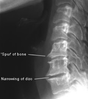
The first one is -osis, so this creates the word spondylosis. Although -osis can denote the “condition of being a …”, in medicine it frequently means a disease or pathological process. Think diverticulosis of the colon. Spondylosis usually denotes a degenerative process of the spine. This is typically due to arthritis and results in bone spurs and disc narrowing. Here is an image of a spine with significant spondylosis:

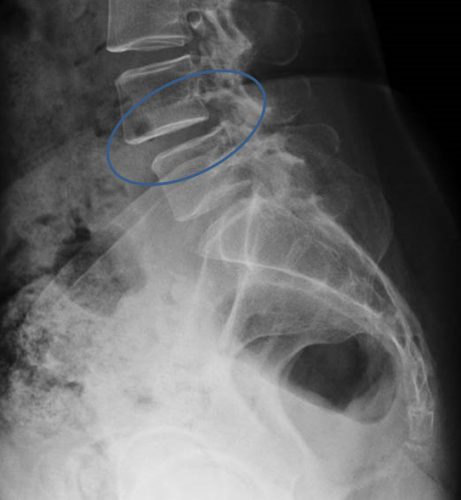
Now let’s add -listhesis. This is another Greek word that means “slipping or falling.” So in this case, the full word means one vertebra slipping over another. Here’s an image of an anterior spondylolisthesis:

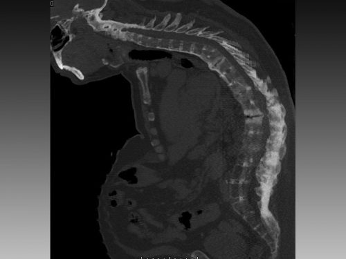
Finally, let’s add -itis. This is the Greek suffix for inflammation. So spondylitis is an inflammatory process of the spine. This can be due to infectious or autoimmune causes. One of the more common types is ankylosis spondylitis, which is an autoimmune variant of rheumatoid arthritis. This causes inflammation of the facet joints and the stabilizing ligaments, leading to fused vertebra and a characteristic patient posture. Here’s a rather extreme case:

I hope this little vocabulary lesson has been helpful. Now go impress your spine specialty colleagues!

Print Friendly, PDF & Email