Category Archives: General

Impact Of Arm Position On Torso CT Scan

CT scan is a valuable tool for initial screening and diagnosis of trauma patients. However, more attention is being paid to radiation exposure and dosing. Besides selecting patients carefully and striving for ALARA radiation dosing (as low as reasonably achievable) by adjusting technique, what else can be done? Obviously, shielding parts of the body that do not need imaging is simple and effective. But what about simply changing body position?

One simple item to consider is arm positioning in torso scanning. There are no consistent recommendations for use in trauma scanning. Patients with arm and shoulder injuries generally keep the affected upper extremity at their side. Radiologists prefer to have the arms up if possible to reduce scatter and provide clearer imaging.

A retrospective review of 710 patients used dose information computed by the CT software and displayed on the console. Radiation exposure was estimated using this data and was stratified by arm positioning. Even though there are some issues with study design, the results were impressive.

There was no difference in scanning time for any arm position. Here are the factoids for radiation dose:

  • Both arms up: 19 mSv (p<0.0000001)
  • Left arm up: 23 mSv
  • Right arm up: 24 mSv
  • Arms down: 25 mSv

Bottom line: Do everything you can to reduce radiation exposure:

  1. Be selective with your imaging. Do you really need it?
  2. Work with your radiologists and physicists to use techniques that reduce dose yet retain image quality
  3. Shield everything that’s not being imaged.
  4. Think hard about getting CT scans in children. They probably don’t need it!
  5. Raise both arms up during torso scanning unless injuries preclude it.

There is a commercial product now available that helps position the arms without tape, paper clips, or other office supply items. It doubles as a pillow for the patient and is held in place by their weight

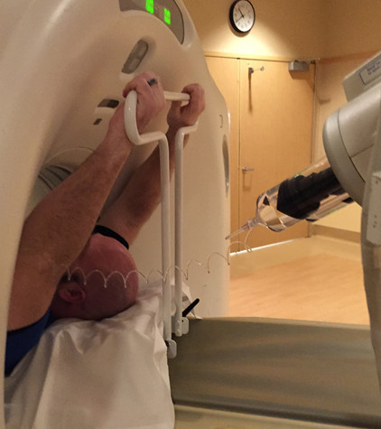
Courtesy of http://accessoryaccommodations.com/

Related posts:

Reference: Influence of arm positioning on radiation dose for whole body computed tomography in trauma patients. J Trauma 70(4):900-905, 2011.

Can Lead Poisoning Occur After A Gunshot?

This is a fairly common question from victims of gunshots and their families. As you know, bullets are routinely left in place unless they are superficial. It may cause more damage to try to extract one, especially if it has come to rest in a deep location. But is there danger in leaving the bullet alone?

One of the classic papers on this topic was published in 1982 by Erwin Thal at Parkland Hospital in Dallas. The paper recounted a series of 16 patients who had developed signs and symptoms of lead poisoning (plumbism) after a gunshot or shotgun injury. The common thread in these cases was that the injury involved a joint or bursa near a joint. In some cases the missile passed through the joint/bursa but came to rest nearby, and a synovial pseudocyst formed which included the piece of lead. The joint fluid bathing the projectile caused lead to leach into the circulation.

The patients in the Parkland paper developed symptoms anywhere from 3 days to 40 years after injury. As is the case with plumbism, symptoms were variable and nonspecific. Patients presented with abdominal pain, anemia, cognitive problems, renal dysfunction and seizures to name a few.

Bottom line: Any patient with a bullet or lead shot that is located in or near a joint or bursa should have the missile(s) promptly and surgically removed. Any lead that has come to rest within the GI tract (particularly the stomach) must be removed as well. If a patient presents with odd symptoms and has a history of a retained bullet, obtain a toxicology consult and begin a workup for lead poisoning. If levels are elevated, the missile must be extracted. Chelation therapy should be started preop because manipulation of the site may further increase lead levels. The missile and any stained tissues or pseudocyst must be removed in their entirety.

Reference: Lead poisoning from retained bullets. Ann Surg 195(3):305-313, 1982.

February Newsletter Update: REBOA

I want everyone to know that you did not miss the newsletter this month! It’s just taking a bit longer than expected to write it because there is so much interesting stuff that’s been written. I’ll have it to subscribers on Feb 28, and post it publicly in the blog the following week.

Subscribe now and be sure to get it first!  So sign up for early delivery now by clicking here!

Pick up back issues here!

Misleading Abstract Alert: Injuries Identified By Chest CT

Here is another one of those papers that have this nicely done abstract that arrives at what seems to be a reasonable conclusion. But then you sit back and think about it. And it’s no longer so reasonable.

This study seems like it should be a good one! It’s a multi-center trial involving data from ten level I trauma centers. The research infrastructure used to collect the data and the statistical analyses for this retrospective review were sound.

Here are the factoids:

  • Of nearly 15,000 patients with blunt chest trauma, about 6,000 (40%) underwent both chest x-ray and CT
  • 25% (1,454) of these patient had new injuries discovered by the CT
  • 954 were truly occult, only being found on the CT; the remaining 500 scans found more injuries than seen on chest x-ray
  • 202 patients had major interventions (chest tube, ventilator, surgery)
  • 343 had minor interventions (admission, extended observation)
  • Chest x-ray was not very good at detecting aortic or diaphragm injury (surprise)
  • 76% of the major interventions were chest tube insertions
  • 32% of of patients with new fractures seen were hospitalized for pain control
  • None of the odds ratios reported were statistically significant

Bottom line: What could possibly go wrong? Ten trauma centers. Six thousand patients. Lots of data points. There are two major issues. First, the primary outcome was a major intervention based on the chest CT. The problem with having so many participating centers is that it is hard to figure out why they performed the interventions. Are they saying that a pneumothorax or hemothorax that was invisible on chest x-ray required a chest tube? Based on whose judgment? Unfortunately, that is a big variable. The authors admit that they did not know whether “interventions based on chest CT were truly necessary or beneficial because we did not study patient outcomes” and that the decisions for intervention “were largely made by residents (usually) or fellows.”

And the secondary outcome was admission or extended observation based on the chest CT. Yet these admissions were primarily for pain management in patients with fractures. Did the patients develop additional pain due to irradiation, or was it there all along?

So adding a chest CT greatly increases the likelihood of doing additional procedures. And it is difficult to tell (from this study) if those procedures were truly necessary. But we know that they can certainly be dangerous. If you back out all of the potentially unnecessary chest tubes and the admissions for pain that should have been admitted anyway, this study demonstrates very little additional value from CT.

A well-crafted imaging guideline will help determine which patients really need CT to identify patients with those occult injuries that are dangerous enough that they can’t be missed. The authors even conclude that “a validated decision instrument to support clinical judgment is needed.”

Related posts:

Reference: Prevalence and clinical import of thoracic injury identified by chest computed tomography but not chesty radiography in blunt trauma: multicenter prospective cohort study. Annals Emerg Med 66(6):589-600, 2015.

The Contaminated Patient

In my last post, I presented the issue of dealing with a surprise patient who was both in arrest and contaminated with gasoline. They are brought into your resuscitation room without warning of the potential hazard. Now that they are here, what do you do?

Thanks for the many online and email responses. This is a tough question, because there are so many variables to think about. And you have to make decisions very quickly. Here’s a rundown on my thought processes.

First, if you get an indication that there might be any type of contamination, insist that your prehospital providers hold the patient outside the ED. Have part of your trauma team waiting at the ambulance dock to do a quick assessment there. Another minute or two of Lucas CPR will not make a difference. Use your best judgment as to how much of a hazard is posed by the fuel/mystery liquid/white powder. But err on the side of being conservative so you don’t end up shutting your entire ED down due to contamination. If in doubt, immediately move to your decontamination area.

If the patient ends up deep in your ED before anyone recognizes that there might be a contamination problem, you must heed three overarching principles:

  • Limit contamination to the rest of your facility. Close the doors to the resuscitation room. Notify security and your hazardous materials team so they can start working on containment and safety issues outside the room. Failure to do this can take your entire hospital offline. If the situation turns out to be a multiple or mass casualty event and your hospital was the only one able to respond, you’ve just created a catastrophe and delayed treatment for the other patients.
  • Ensure the safety of your team. This is a great reason to require and enforce that everyone on the team dress up completely for every resuscitation. You never know where your patients have been, and when one of these will sneak in.
  • Continuously assess the risk:benefit ratio. Is the contamination a minor irritant? What is the danger to the team? The ED/hospital? How likely are your efforts to save the patient to succeed? As soon as the ratio goes bad, rethink the options and act accordingly.

Bottom line: In situations like this, think fast and think globally. Don’t just consider the patient. There may be many more lives at stake, and this can and should factor into your decisions about where and how long to continue resuscitation.

In this case, we were certain it was only gasoline. We closed the doors and quickly stripped the patient, bagging the clothes tightly. We tried not to generate any sparks, but we are surrounded by all kinds of electrical equipment. Defibrillation was out of the question. After the event was finished, it was time to wash everything down and start thinking about what would have happened if this had been something more toxic than fuel!

Related post: