Category Archives: General

Autopsy Reports and Performance Improvement

Autopsy reports have traditionally been used as part of the trauma performance improvement (PI) process. They are typically a tool to help determine preventability of death in cases where the etiology is not clear. Deaths that occur immediately prior to arrival or in the ED are typically those in which most questions arise.

The American College of Surgeons Trauma Verification Program includes a question on what percentage of deaths at a trauma center undergo autopsy. Low numbers are usually discussed further, and strategies for improving them are considered. But are autopsies really that helpful?

A total of 434 trauma fatalities in one state over a one year period were reviewed by a multidisciplinary committee and preventability of death was determined. Changes in preventability and diagnosis were noted after autopsy results were available. 

Here are the factoids:

  • The autopsy rate was 83% for prehospital deaths and 37% for in-hospital deaths 
  • Only 69% were complete autopsies; the remainder were limited internal or external only exams
  • Addition of autopsy information changed the preventability determination in 2 prehospital deaths and 1 in-hospital death (1%)
  • In contrast to this number, it changed the cause of death in about 40% of cases, mostly in the prehospital deaths

Bottom line: From a purely performance improvement standpoint, autopsy does not appear to add much to determining preventability of death. It may modify the cause of death, which could be of interest to law enforcement personnel. And it may modify some of the diagnoses recorded in the trauma registry. I would still recommend obtaining the reports for their educational value, especially for those of you who are part of residency training programs.

Related post:

Reference: Dead men tell no tales: analysis of the utility of autopsy reports in trauma system performance improvement activities. J Trauma 73(3): 587-590, 2012.

CT Cystography For Bladder Trauma

Bladder injury after blunt trauma is relatively uncommon, but needs to be identified promptly. Nearly every patient (97%+) with a bladder injury will have hematuria that is visible to the naked eye. This should prompt the trauma professional to obtain a CT of the abdomen/pelvis and a CT cystogram.

The CT of the abdomen and pelvis will identify any renal or ureteral (extremely rare!) source for the hematuria. The CT cystogram will demonstrate a bladder injury, but only if done properly!

During most trauma CT scanning of the abdomen and pelvis, the bladder is allowed to passively fill, either by having no urinary catheter and having the patient hold it, or by clamping the catheter if it is present. Unfortunately, this does not provide enough pressure to demonstrate small intraperitoneal bladder injuries and most extraperitoneal injuries.

The proper technique involves infusing contrast into the bladder through a urinary catheter. At least 350cc of dilute contrast solution must be instilled for proper distension and accurate diagnosis. This can be done prior to the abdominal scan. Once the initial scan has been obtained, the bladder must be emptied and a focused scan of just the bladder should be performed (post-void images). Several papers have shown that this technique is as accurate as conventional retrograde cystography, with 100% sensitivity and specificity for intraperitoneal ruptures. The sensitivity for extraperitoneal injury was slightly less at 93%.

Bottom line: Gross hematuria equals CT of the abdomen/pelvis and a proper CT cystogram, as described above. Don’t try to cheat and passively fill the bladder. You will miss about half of these injuries!

Related posts:

Reference: CT cystography with multiplanar reformation for suspected bladder rupture: experience in 234 cases. Am J Roentgenol 187(5):1296-302, 2006.

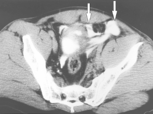
Intraperitoneal bladder rupture

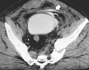
Extraperitoneal bladder injury

Practical Tip: Evaluation of Hematuria in Blunt Trauma

Bloody urine is a relatively uncommon finding in blunt trauma patients. Hematuria ranges from microscopic to gross. Microscopic means blood that can only be seen with a microscope, and gross means visible to the naked eye. In trauma, we only care about gross hematuria, which ranges from the faintest of pink to the deepest red.

In the picture above gross hematuria is present in all tubes but the far right one. Those four will need further evaluation.

In trauma, gross hematuria is a result of an injury to kidney, ureter or bladder. Blunt injury to the ureter is so rare it’s reportable, so you can pretty much forget that one unless the mechanism is extreme. So you really just need to focus on kidney and bladder.

Any victim of blunt trauma that presents with visible hematuria needs to be evaluated by CT of the abdomen and pelvis with an added CT cystogram. Standard CT technique is done without a urinary catheter, or with the catheter clamped. This is not acceptable for hematuria evaluation, as only 50% of bladder injuries show up with this technique.

CT cystogram is an add-on to the standard CT, and consists of the administration of contrast into the bladder which is then kept under pressure while the scan is performed. Delayed slices through the pelvis after the bladder is depressurized and emptied is routine. Nearly 100% of bladder injuries are detected using this technique.

If the CT shows a renal laceration or hematoma, the patient should be admitted and managed according to your solid organ injury protocol. Kidney injuries fare better that livers and spleens, and only rarely require surgery. If no kidney or bladder injury is seen, the default diagnosis of a renal contusion is the culprit. No treatment is needed, and the patient can be discharged if no other injuries are present. The blood will clear over a few days, but may disappear and reappear a few times in the process. Be sure to warn the patient that this may occur, or you may receive some surprise phone calls. The patient can followup with their primary care physician in a week or two.

The majority of these injuries do not require urologic consultation. Complex injuries with extravasation of urine out of the kidney, or injuries to the collecting system should be referred to a urologist, however.

Trauma MedEd Newsletter: REBOA!

The March newsletter is now available! Click the link below to download. 

The newest, hottest thing these days seems to be REBOA. Curious? This issue explores the things you always wanted to know about it.

In this issue you’ll find articles on:

  • What Is REBOA?
  • Who is REBOA For?
  • How Is REBOA Performed?
  • What Are The Results For REBOA?
  • What’s The Bottom Line?

Subscribers received the newsletter last Sunday . If you want to subscribe to get early delivery in the future (and download back issues), click here.

Click here to download newsletter.

Bedrest After Pediatric Liver/Spleen Injury? Really?

A set of guidelines for management of blunt solid organ injury in children developed by the American Pediatric Surgical Association was originally published in 1999. One of the elements of the guideline was to place the child on bedrest for a period of time after the injury. Arbitrarily, this period was defined as the injury grade plus one day. So for a grade 3 spleen injury, the child would have to stay in bed for 4 days (!).

A paper published in 2013 looked at the impact of shortening this time interval. Over a 6 year period, all pediatric liver and spleen injuries from blunt trauma were identified and an abbreviated bedrest protocol was implemented. For low grade injuries (grade 1-2), children were kept in bed for 1 day, and for higher grade injuries this was extended to 2 days.

Here are the factoids:

  • 249 patients were enrolled (about 40 per year) with an average age of 10. “Bedrest was applicable for 199 patients, 80%.” Huh? Does that mean that 50 patients were excluded due to surgeon preference?
  • The organ injured was about 50:50 for spleen vs liver. Twelve children injured both.
  • Mean injury grade was 2.7, which is fairly high
  • Mean bedrest was 1.6 days, and mean hospital stay was 2.5
  • Bedrest was the limiting factor for hospital stay in 62% of cases
  • There were no delayed complications of the injury

Bottom line: Come on! Most centers don’t keep adult patients at bedrest this long, and we learned about solid organ injury management from kids! Children almost never fail nonop management, so why treat them more restrictively than adults? And have you ever tried to keep a child at bedrest? Impossible! This study is too underpowered to give real statistically valid results, but it certainly paints a good picture of what works. We recently updated our adult and pediatric protocols to eliminate bedrest and npo status. Let’s get rid of these anachronisms once and for all!

Reference: Follow up of prospective validation of an abbreviated bedrest protocol in the management of blunt spleen and liver injury in children. J Ped Surg 48(12):2437-2441, 2013.