Category Archives: General

What The Heck? Head CT – The Answer

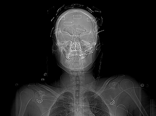
It appears that no one was able to figure this one out! To recap, a young person (female in this case) sustained blunt trauma to the head. When her head was scanned, the following was found on the scout scan:

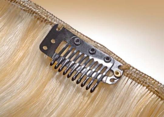
What is all this odd stuff? They look like some kind of metallic clips placed all over the head. The answer? Hair extensions! Here’s what they look like up close.

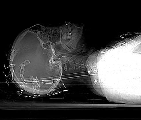
Unfortunately, they cannot be left in place during the CT. The amount of scatter is significant enough to degrade the quality of the study. By definition, if you have taken your patient to CT, they are stable and you have a little time. So carefully remove all of the extensions and place them in a bag and save them for the patient (they can be expensive!). 

Make sure the CT tech obtains another topogram to confirm removal of all of the extensions. Then proceed with your CT as usual!

Print Friendly, PDF & Email

Why Do They Call It: Extra-axial Blood?

You’ve seen it on head CT reports. “The patient has a collection of extra-axial blood…” Then it goes on to describe the location and size of a subdural hematoma. But why is it called “extra-axial?”

The answer lies in the embryology of the central nervous system. Yes, it’s been a long time since any of us have read anything about that. Early animals had a straight neural tube, which slowly evolved into a brain and spinal cord. This is known as the axis of the nervous system.

The brains of early vertebrates developed at the end of the neural tube, and were oriented in the same longitudinal axis as the rest of it. As brains got bigger, a 90 degree bend occurred at the cephalic flexure.

So in humans, there is a difference between the body axis and the brain axis. But the brain axis is what really counts. This means that any blood outside of the brain axis is defined as extra-axial.

Bottom line: Extra-axial blood is defined as any bleeding outside of the brain parenchyma. This includes subdural and epidural hematomas, and subarachnoid hemorrhage. It does not include any intraparenchymal bleeding like contusions, strokes, or hematomas.

Print Friendly, PDF & Email

What’s The Best Test For Blunt Cerebrovascular Injury?

Blunt injury to the carotids or vertebrals (BCVI) is a little more common than originally thought, affecting about 1% of blunt trauma patients. We have many tools available to help us diagnose the problem: duplex ultrasound, CT angiography (CTA), MR angiography (MRA), and even good old conventional 4 vessel angiography

But which one is “best?” This is a tough question, because there is always some interplay between clinical accuracy and cost. The surgical group at the Medical College of Wisconsin – Milwaukee did a nice job teasing some answers from existing literature on the topic. The authors tried to take a comprehensive look at costs, including money spent to prevent stroke, the cost of complications of therapy, and the overall cost to society if the patient suffers a stroke.

Here are the factoids:

  • For patients at risk for BCVI, the stroke rate is 11% without screening, 6% with duplex ultrasound screening, 4% with MRA, and 1% with either CTA or conventional angiography
  • From a societal standpoint (includes the lifetime costs of stroke for the patient), CTA is the most cost effective at $3,727 per patient
  • From the hospital standpoint (does not include lifetime cost), no screening is the most cost effective, but has the highest stroke rate (11%)
  • CTA prevents the most strokes, and costs about $10,000 per patient while decreasing societal costs by about $32,000 per patient screened

Bottom line: The “best” test for patients at risk for blunt cerebrovascular injury is the CT angiogram. It minimzes the stroke rate, and provides information on all four vessels supplying the brain, which is probably why the duplex ultrasound has a higher miss rate (can’t see the vertebrals or into the skull). But how do you decide who is at risk for this problem. Tune in tomorrow!

Reference: Screening for Blunt Cerebrovascular Injuries is Cost-Effective. J Trauma 70(5):1051-1057, 2011.

Print Friendly, PDF & Email

Procedural Complications: Residents vs Advanced Practice Providers

With the implementation of resident work hour restrictions 10 years ago, resident participation in clinical care has declined. In order to make up for this loss of clinical manpower and expertise, many hospitals have added advanced clinical providers (ACPs, nurse practitioners and physician assistants). These ACPs are being given more and more advanced responsibilities, in all clinical settings. This includes performing invasive procedures on critically ill patients.

A recent study from Carolinas Medical Center in Charlotte NC compared complication rates for invasive procedures performed by ACPs vs residents in a Level I trauma center setting.

A one year retrospective study was carried out. Here are the factoids:

  • Residents were either surgery or emergency medicine PGY2s
  • ACPs and residents underwent an orientation and animal- or simulation-based training in procedures
  • All procedures were supervised by an attending physician
  • Arterial lines, central venous lines, chest tubes, percutaneous endoscopic gastrostomy, tracheostomy, and broncho-alveolar lavage performances were studied
  • Residents performed 1020 procedures and had 21 complications (2%)
  • ACPs performed 555 procedures and had 11 complications (2%)
  • ICU and hospital length of stay, and mortality rates were no different between the groups

Bottom line: Resident and ACP performance of invasive procedures is comparable. As residents become less available for these procedures, ACPs can (and will) be hired to  take their place. Although this is great news for hospitals that need manpower to assist their surgeons and emergency physicians, it should be another wakeup call for training programs and educators to show that resident education will continue to degrade.

Reference: Comparison of procedural complications between resident physicians and advanced clinical providers. J Trauma 77(1):143-147, 2014.

Print Friendly, PDF & Email