Category Archives: General

Resident Work Hour Restrictions And Neurotrauma Complications

In the US, resident work hour restrictions went into effect in 2003, limiting the total number of hours worked per week and the number of consecutive hours without a break. The idea was that fatigue caused errors, which translates into patient complications or worse. Has this panned out? A number of previous publications have found no change; only a few have shown some benefit.

Researchers at Massachusetts General Hospital decided to apply the acid test to this theory. They selected a group of patients who were critically ill and challenging to care for, taken care of by a group of residents who had long work hours and were involved in long operative cases. The AHRQ National Inpatient Sampling Database was studied, comparing the outcomes of neurotrauma patients before and after work hours were initiated and in teaching and non-teaching centers.

A huge number of records were analyzed (40,000 before work hours restrictions, 67,000 after). The findings were intriguing:

  • The overall complication rate was the same before and after restrictions (1.2%)
  • The complication rate was 25% higher in teaching hospitals after restrictions took effect. It appears that this also correlated with higher hospital charges after restrictions.
  • Logistic regression was used to figure out whether this difference was from duty hours or just from the involvement of residents in care. Only duty hours were significant in this analysis.
  • If injury severity was included in the analysis, there were no differences in complications at all
  • There were no differences in mortality rates between any of the groups

Bottom line: Yes, fatigue is bad (see my previous posts below). But here is another (correlation) study that doesn’t bear out the original reasons to restrict resident work hours. In actuality, complications and charges increased after the restrictions went into effect. It is possible that the checks and balances in the system were effective in protecting patients from adverse outcomes. Could the changes in this study be due to staffing changes to meet the restrictions, which results in chronic understaffing which undercuts those checks and balances? Studies of this type can’t tell us that. And unfortunately, restrictions in the US are not going to go away, they’ll probably get worse.

Related posts:

Reference: Higher Complications and No Improvement in Mortality in the ACGME Resident Duty-Hour Restriction Era: An Analysis of More Than 107?000 Neurosurgical Trauma Patients in the Nationwide Inpatient Sample Database. Neurosurgery 70(6):1369-1382, 2012.

Print Friendly, PDF & Email

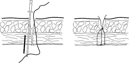
Technique: How To Close A Full Thickness Stab (Abdomen) Laparoscopically

The algorithm for evaluating a stab to the anterior abdomen includes a number of different techniques for evaluation. In some cases where the chance of entry into the abdomen is thought to be low probability, endoscopic exploration can be used. What if a full thickness stab is detected, but the surgeon is able confirm that no abdominal injuries are present? Should the stab defect be closed?

There is no good data that tells us the incidence of ventral hernia from stab wounds. We do know that 10mm endoscopic port sites and larger can be the source of a ventral hernia and possible bowel obstruction after laparoscopic surgery, so it stands to reason (but be careful) that the same thing could happen with larger stabs. So why not close them?

A number of commercial devices have been developed for port site closure during endoscopic surgery (Carter Thomason Closure System, Cooper Surgical; Endo Close, Covidien). A group in Tokyo published a description of the technique using the former device to close the fascial defect of a self-inflicted stab wound.

Bottom line: This is an interesting use for a device used for closing more controlled stab wounds (surgical port sites) in less controlled ones. It seems fair to extrapolate our current experience from laparoscopic surgery to trauma in this case. I would be very interested to hear from anyone who is currently using this technique.

Reference: A quick and easy closure technique for abdominal stab wound after diagnostic laparoscopy. J Trauma 72(5):1448-1449, 2012.

Print Friendly, PDF & Email

Angioembolization For Splenic Injury

Initial nonoperative management of splenic injury is standard in hemodynamically stable patients. Over the past decade, the success rates have climbed by adding angioembolization to the algorithm, according to several published series. However, the objective benefit and specific indications have not been worked out.

A paper published this month by the University of Florida, Jacksonville used the NTRACS registry to try to clarify these issues. They identified 1039 patients undergoing nonoperative management (NOM) over a nearly 10 year period. Patients who died shortly after arrival, those who went directly to OR for hemodynamic reasons, and children were excluded, leaving 539 patients. Only about 1/6 of the patients underwent embolization. 

The overall failure rate was about 4%, a little higher in the non-angio patients, a little lower with angio. Incidentally, the angio group had significantly higher injury severity (26 vs 20). Analysis of the lower grade spleen injury group showed no improvement in success rate by adding angio. However, the high grade groups (grades IV-V) did benefit by adding this procedure. Similarly, success improved when performing angio in patients with contrast blush or evidence of slow, ongoing bleeding. If NOM did fail, it usually occurred on day 2.

Bottom line: Although we’ve been adding angio to non-operative management of spleen and liver injury for a decade, here’s the first paper that has been able to define the real indications for doing it. First, all unstable patients go to the OR (don’t even consider nonop management). In the remaining patients, if the CT shows a grade IV or V injury, or a contrast blush, angio is recommended. If neither of these is noted, but the hemoglobin continues to decline “too quickly” (surgeon judgement), then a trip to angio is also warranted. Applying these principles can increase your success rate to about 96%.

Related post:

Reference: Selective angiographic embolization of blunt splenic traumatic injuries in adults decreases failure rate of nonoperative management. J Trauma 72(5):1127-1134, 2012.

Print Friendly, PDF & Email

Lateral Canthotomy For Orbital Compartment Syndrome

I’ve previously written about the orbital compartment syndrome and described the symptoms in the awake patient. I’d like to share a video of the procedure and provide a tip for diagnosing it in unconscious patients.

Patients at risk will have obvious facial trauma. During your physical exam, look for proptosis. This is caused by swelling or hemorrhage behind the globe pushing it forward. It may manifest itself as uneven opening of the eyelids, with the affected side being “propped” open (get it?). But in trauma, there may be significant edema which keeps the lid closed.

The easiest way to observe proptosis is to stand above the patients head, looking toward their feet. Crouch down so you can look across the lids in a direction horizontal to the floor. You should easily be able to detect if one eye protrudes further that the other.

You can also do a poor man’s compartment pressure test by gently using your thumbs to compress both globes simultaneously. If there is substantial difference in resistance between the two sides, a compartment syndrome may be present. Important note! Do a thorough globe exam first! If there is any evidence of globe rupture (hyphema, irregular pupil, extra tissue in the anterior compartment), don’t press the eye or perform a canthotomy. CT scan of the facial bones can help confirm the diagnosis if a mass effect is seen in the orbit or if the optic nerve appears to be on stretch.

The orbital compartment syndrome is an emergency! Once diagnosed, immediately proceed to canthotomy. Otherwise, damage to the optic nerve and retina is likely, and the patient may become blind in the affected eye.

Related post:

Print Friendly, PDF & Email