Category Archives: General

Why Do Trauma Patients Get Readmitted?

Readmission of any patient to the hospital is considered a quality indicator. Was the patient discharged too soon for some reason? Were there any missed or undertreated injuries? Information from the Medicare system in the US (remember, this represents an older age group than the usual trauma patient) indicates that 18% of patients are readmitted and 13% of these are potentially preventable.

A non-academic Level II trauma center in Indiana retrospectively reviewed their admissions and readmissions over a 3 year period and excluded patients who were readmitted on a planned basis (surgery), with a new injury, and those who died. This left about 5,000 patients for review. Of those, 98 were identified as unexpected readmissions. 

There were 6 major causes for readmission:

  • Wound (23) – cellulitis, abscess, thrombophlebitis. Two thirds required surgery, and 4 required amputation. All of these amputations were lower extremity procedures in obese or morbidly obese patients.
  • Abdominal (16) – ileus, missed injury, abscess. Five required a non-invasive procedure (mainly endoscopy). Only 2 required OR, and both were splenectomy for spleen infarction after angioembolization.
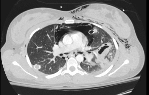
  • Pulmonary (7) – pneumonia, empyema, pneumothorax, effusion. Two patients required an invasive procedure (decortication, tube placement).
  • Thromboembolic (4) – DVT and PE.  Two patients were admitted with DVT, 2 with PE, and 1 needed surgery for a bleed due to anticoagulation.
  • CNS (21) –  mental status or peripheral neuro exam change. Eight had subdural hematomas that required drainage; 3 had spine fractures that failed nonoperative management. 
  • Hematoma (5) – enlargement of a pre-existing hematoma. Two required surgical drainage.

About 14% of readmissions were considered to be non-preventable by a single senior surgeon. Wound complications had the highest preventability and CNS changes the lowest. Half occurred prior to the first followup visit, which was typically scheduled 2-3 weeks after discharge. This prompted the authors to change their routine followup to 7 days. 

Bottom line: This retrospective study suffers from the usual weaknesses. However, it is an interesting glimpse into a practice with fewer than the usual number patients lost to followup. The readmission rate was 2%, which is pretty good. One in 7 were considered “preventable.” Wounds and pulmonary problems were the biggest contributors. I recommend that wound and pulmonary status be thoroughly assessed prior to discharge to bring this number down further. Personally, I would not change the routine followup date to 1 week, because most patients have far more complaints that are of little clinical importance than compared to 2 weeks after discharge.

Reference: Readmission of trauma patients in a nonacademic Level II trauma center. J Trauma 72(2):531-536, 2012.

Print Friendly, PDF & Email

Can Lead Poisoning Occur After A Gunshot?

This is a fairly common question from victims of gunshots and their families. As you know, bullets are routinely left in place unless they are superficial. It may cause more damage to try to extract one, especially if it has come to rest in a deep location. But is there danger in leaving the bullet alone?

One of the classic papers on this topic was published in 1982 by Erwin Thal at Parkland Hospital in Dallas. The paper recounted a series of 16 patients who had developed signs and symptoms of lead poisoning (plumbism) after a gunshot or shotgun injury. The common thread in these cases was that the injury involved a joint or bursa near a joint. In some cases the missile passed through the joint/bursa but came to rest nearby, and a synovial pseudocyst formed which included the piece of lead. The joint fluid bathing the projectile caused lead to leach into the circulation.

The patients in the Parkland paper developed symptoms anywhere from 3 days to 40 years after injury. As is the case with plumbism, symptoms were variable and nonspecific. Patients presented with abdominal pain, anemia, cognitive problems, renal dysfunction and seizures to name a few.

Bottom line: Any patient with a bullet or lead shot that is located in or near a joint or bursa should have the missile(s) promptly and surgically removed. Any lead that has come to rest within the GI tract (particularly the stomach) must be removed as well. If a patient presents with odd symptoms and has a history of a retained bullet, obtain a toxicology consult and begin a workup for lead poisoning. If levels are elevated, the missile must be extracted. Chelation therapy should be started preop because manipulation of the site may further increase lead levels. The missile and any stained tissues or pseudocyst must be removed in their entirety.

Reference: Lead poisoning from retained bullets. Ann Surg 195(3):305-313, 1982.

Print Friendly, PDF & Email

How Long Should We Watch Intracerebral Hemorrhage?

Patients with traumatic brain injury (TBI) severe enough to cause bleeding are usually admitted to the hospital for observation and in many cases, repeat CT scanning. Those with small intracranial hemorrhages (ICH) may experience progression of the bleeding, and a small percentage of cases may need operative intervention (1-3%). Questions we typically face are, how long should we watch for progression, and how often should we scan?

A retrospective cohort study was carried out at UMD-NJ, looking for answers for a specific subset of these patients. Specifically, they had to have a mild blunt TBI (loss of consciousness and/or retrograde amnesia, GCS in the ED of 13-15) and a positive head CT. They classified any type of hemorrhage into or around the brain as positive. 

During a 3 year period, 474 adults were enrolled but only 341 were eligible for the study. They were excluded due to previous injury, presence of a mass (not trauma), need for immediate neurosurgical intervention, or failure to get a second CT scan. The authors found:

  • 7% of patients were taking anticoagulants! This is surprisingly high. Interestingly, 15 were subtherapeutic, 3 were therapeutic and 2 were supratherapeutic.
  • Subarachnoid hemorrhage was the most common finding on CT (54%). Intraparenchymal hemorrhage was next most common (48%) Many patients had more than one type of bleed.
  • The injury worsened between the first and second scans in 31% of patients. This number increased to 46% in patients taking anticoagulants. 
  • About 97% of bleeds stopped progressing by 24 hrs post-injury.

Bottom line: Most centers are probably overdoing the observation and repeat scan thing. More than two thirds of bleeds are stable by the first scan (first and second scans identical), and nearly all stop progressing within 24 hours. It’s very likely that patients who are not on anticoagulants and who have a stable neuro exam and stable symptoms can get just one scan and 24 hours of observation. Persistent headache, nausea, failure to ambulate well, or other symptoms warrant a repeat scan and longer observation.

Related posts:

Reference: The temporal course of intracranial haemorrhage progression: How long is observation necessary? Injury 43(12):2122-2125, 2012.

Print Friendly, PDF & Email

Table Of Contents Updated!

Looking for specific content from the Trauma Professional’s Blog? I’ve posted almost 600 items over the last 3 years here, so there’s a lot of stuff to sift through! There are several ways to do it, and here are some tips.

If you look over in the column to the right, you’ll see three tools to help you:

  • The search box. Type in a search term, and Google will do a nice search of just the blog. Note: older versions of Internet Explorer don’t work quite right, so you have to actually click the Search button. Hitting enter will do a Google search of the whole internet, not just the blog.
  • The Indexed archive button. This is a categorized list of all the content on this blog. It’s getting long! I’m in the process of streamling it so it’s more manageable.
  • The Tumblr archive button. This presents a nice thumbnail view of each month’s posts. You can look at all the recent titles, but it’s not really very searchable.
  • The Submit Your Request button. This one is the best! Don’t see what you’re looking for? Send me a request! You’re welcome to email or Skype me as well! I get some of my best material from requests. If you’re curious about a specific topic, other people probably are as well!

Print Friendly, PDF & Email

Answer: What The Heck Video! Part 2

This patient was involved in a motor vehicle crash with significant chest trauma. They’ve been intubated and are oxygenating and ventilating well. What to do next?

First, the endotracheal tube was a bit deep, which can create its own problems. It was pulled back a few centimeters. Since the patient was hemodynamically stable, a CT angio of the chest would be very helpful to try to figure out the pathology. Here’s a representative slice from the scan.

There are a few striking findings here:

  • Extensive subcutaneous emphysema
  • Large pneummediastinum around the heart
  • Significant injury to the left lung (note the pneumatocele, an air filled collection)
  • Atelectasis of the left lung despite repositioning of the ET tube

The combination of of the above is highly suggestive of a large airway injury. Since the entire lung was affected, it is most likely a mainstem bronchus injury. Usually, these are accompanied by a large air leak from the chest tube, but not in this case.

This prompted the bronchoscopy shown two days ago. The image is oriented such that the left mainstem bronchus was on the right side of the video. A bronchial tear is visible on the lateral aspect, just before the takeoff of the upper lobe bronchus. You can get the impression of a beating heart beating somewhere nearby. And when the camera pops through the laceration, you can actually see the thoracic aortic coursing away toward the diaphragm!

Related post:

Print Friendly, PDF & Email