Category Archives: General

Syncope Workup in Trauma Patients

Syncope accounts for 1-2% of all ED visits, and is a factor in some patients with blunt trauma, especially the elderly. If syncope is suspected, a “syncope workup” is frequently ordered. Just what this consists of is poorly defined. Even less understood is how useful the syncope workup really is.

Researchers at Yale retrospectively looked at their experience doing syncope workups in trauma patients. They were interested in seeing what was typically ordered, if it was clinically useful, and if it impacted length of stay. 

A total of 14% of trauma patients had syncope as a possible contributor to their injury. The investigators found that the following tests were typically ordered in these patients:

  • Carotid ultrasound (96%)
  • 2D Echo (96%)
  • Cardiac enzymes (81%)
  • Cardiology consult (23%)
  • Neurology consult (11%)
  • EEG (7%)
  • MRI (6%)

Most of this testing was normal. About 3% of cardiac enzymes were abnormal, as were 5% of carotid imaging and 4% of echocardiograms. 

Important! Of the patients who underwent an intervention after workup, 69% could have been identified based on history, physical exam, or EKG and did not depend on any of the other diagnostic tests.

Conclusion: Syncope workup is not needed routinely in trauma patients with syncope as a contributing factor. Need for intervention can usually be determined by history, exam and EKG performed in the ED. In this study, $216,000 in excess costs would have been saved!

Reference: Routine or protocol evaluation of trauma patients with suspected syncope is unnecessary. J Trauma 70(2):428-432, 2011.

Print Friendly, PDF & Email

Pop Quiz – Final Answer

Here’s some background info to go with the xray presented Wednesday:

  • Thin
  • Athlete
  • Epigastric trauma

Put these three together and you get a patient engaged in significant physical activity who was struck in the abdomen. If no pads are involved in the sport, the patient has little padding of their own.

This is a setup for pancreatic or duodenal trauma. This patient presented after being struck in the epigastrium by an elbow during a soccer game. It hurt, but wasn’t bad enough to stop playing. The following day, she was a little sore but felt bloated and started throwing up after breakfast.

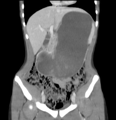
In the ED, a CT was obtained. Here is a coronal view showing the distended stomach:

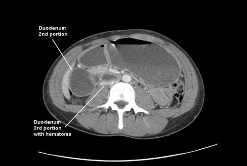
Axial views showed obstruction in the proximal 3rd portion of the duodenum, right over the spine:

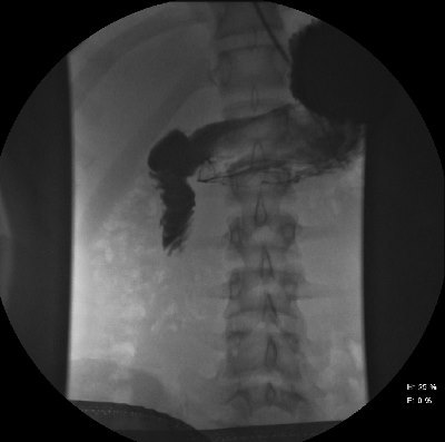
An (unnecessary) contrast study was performed, which confirmed the pathology. Note the tapering and corkscrew appearance of the duodenal folds.


Final diagnosis: duodenal hematoma. This is a crushing injury from compression of the anterior abdominal wall against the spine. The third portion of the duodenum lies over the spine, as does the pancreas, so both are likely to be injured. The latter organ appeared normal on the CT.

Management of blunt duodenal hematoma is simple: wait on it. These will generally resolve quickly over the course of a few days. NG decompression is mandatory, since nothing will pass the obstructed area (saliva, gastic juice, and pancreatic effluent, which add up to 2L+ of fluid per day). In rare cases, parenteral nutrition may be needed if resolution time is approaching the one week mark or in smaller children. A surgical approach with drainage of the hematoma has a low but significant morbidity compared to just waiting. Athletes may return to play soon after recovery.

Print Friendly, PDF & Email

Trauma Mythbusters: NSAIDs And Fracture Healing

Trauma hurts like hell. Over the years, we’ve developed quite a few ways of combating this pain. A number of drug classes have been developed to reduce it. One of the more common non-narcotic drug classes are the NSAIDs. As I’ve mentioned before, every drug has dozens of effects. Drug companies market a particular medication based on one of the predominant effects. All the others are considered side effects.

NSAIDs are not unique; they have lots of side effects as well. In 2003, several papers brought to light possible interactions between these drugs and fracture healing. Specifically, there were questions about these drugs interfering with the healing process and of increasing the number of delayed unions or nonunions. But once again, how convincing were these papers, really?

It would seem to make sense that NSAIDs could interfere with bone healing. This  process relies heavily on the regulation of osteoblast and osteoclast function, which itself is regulated by prostaglandins. Since prostaglandins are synthesized by the COX enzymes, COX inhibitors like the NSAIDs should have the potential to impair this process. Indeed, animal studies in rats and rabbits seem to bear this out.

But as we have seen before, good animal studies don’t always translate well to human experience. Although a study from 2005 suggested that NSAID administration in older patients within 90 days of injury had a higher incidence of fracture nonunion, the study design was not a very good one. It is equally likely that patients who required these drugs in this age group may have been at higher risk for nonunion in the first place.

In fact, there are no large, prospective randomized studies that have explored the effect of short-term or long-term NSAID administration on fracture repair. But there have been several smaller studies that showed absolutely no effect on nonunion with short-term administration of this drug class. Yet the dogma that leads us to avoid giving these drugs persists.

Bottom line: Once again, the animal data is clear but the human data is not. Although there are theoretical concerns about their use, there is not enough solid risk:benefit information to abandon short-term NSAID use in patients who really need them. NSAIDs can and should be prescribed in patients with short-term needs and simple fractures.

References:

  1. Effects of nonsteroidal anti-inflammatory drugs on bone formation and soft-tissue healing. J AM Acad Orthop Surg 12:139-43, 2004.
  2. Effect of COX-2 on fracture-healing in the rat femur. J Bone Joint Surg Am 86:116-123, 2004.
  3. Effects of perioperative anti-inflammatory and immunomodulating therapy on surgical wound healing. Pharmacotherapy 25:1566-1591, 2005.
  4. Pharmacological agents and impairment of fracture healing: what is the evidence? Injury 39:384-394, 2008.
  5. High dose nonsteroidal anti-inflammatory drugs compromise spinal fusion. Can J Anaesth 52:506-512, 2005.
Print Friendly, PDF & Email

What To Do? Chest Tube Repositioning

So you’re faced with a chest tube that “someone else” inserted, and the followup chest xray shows that the last drain hole is outside the chest. What to do?

Well, as I mentioned, there is very little written on this topic, just dogma. So here are some practical tips on avoiding or fixing this problem:

  • Don’t let it happen to you! When inserting the tube, make sure that it’s done right! I don’t recommend making large skin incisions just to inspect your work. Most tubes can be inserted through a 2cm incision, but you can’t see into the depths of the wound. There are two tricks: 
  1. In adults with a reasonable BMI, the last hole is in when the tube markings show 12cm (bigger people need bigger numbers, though)
     
  2. After insertion, get into the habit of running a finger down the radiopaque stripe on the tube all the way to the chest wall. If you don’t feel a hole (which is punched through the stripe), this will confirm that the it is inside, and that the tube actually goes into the chest. You may laugh, but I’ve seen them placed under the scapula. This even looks normal on chest xray!
  • Patient with a high BMI may not need anything done. The soft tissue will probably keep the hole occluded. If there is no air leak, just watch it.
     
  • If the tube was just put in and the wound has just been prepped and dressed, and the hole is barely outside the rib line, you might consider repositioning it a centimeter or two. Infection is a real concern, so if in doubt, go to the next step.
     
  • Replace the tube, using a new site. Yes, it’s a nuisance and requires more anesthetic and possibly sedation, but it’s better than treating an empyema in a few days.

Related posts:

Print Friendly, PDF & Email