Category Archives: General

Outcome After Blunt Cerebrovascular Injury (BCVI)

Blunt injuries to the carotid and vertebral arteries are not as uncommon as we used to think. Unfortunately, there’s a lot of controversy surrounding everything about them: screening, management, and outcome. A paper just out detailed outcomes in a (relatively) large series of these patients. 

As expected with this rare injury, it’s a retrospective study. A busy Level I center identified 222 patients with 263 BCVIs over a 4 ½ year period. Twenty four died before discharge and 11 afterwards. Of the remaining patients, only 74 could be located and only 68 could be persuaded to complete an interview and evaluation of their functional status. Functional Independence and Functional Activity Measurements were assessed (FIM/FAM).

Pertinent findings were:

  • 8 patients suffered a stroke during their initial hospital stay (5 were present on arrival in the ED)
  • 5 additional patients had a stroke after discharge
  • Only 20% reached the maximum FIM/FAM scores, even including patients who did not have a stroke
  • Patients with stroke had a significantly lower FIM/FAM
  • There was no difference in FIM/FAM in patients with carotid vs vertebral injury

Bottom Line: Even though it is limited, this is one of the best studies we will see on BCVI because it’s an uncommon problem at most centers. The most important fact here is that the stroke rate was 19% despite discharge on antiplatelet or anticoagulant medications. And if stroke occurs, it causes significant functional problems, as expected. It’s critically important that this injury be screened and identified appropriately, then given appropriate prophylaxis. More on this tomorrow.

Related posts:

Reference: Functional outcomes following blunt cerebrovascular injury. J Trauma 74(4):955-960, 2013.

Print Friendly, PDF & Email

Complications of Splenic Embolization for Trauma

Angioembolization has become a common procedure that can increase the likelihood of success for nonoperative management for splenic trauma. It does have its own set of complications to be aware of, however.

The most obvious complication is mechanical injury to the femoral artery. This occurs in 1 to 3% of patients. It is more common in the very young (small caliber artery) and the elderly (arteries of stone). Rarely, the substance or device that is used for the embolization may migrate or end up on the wrong spot, infarcting something important.

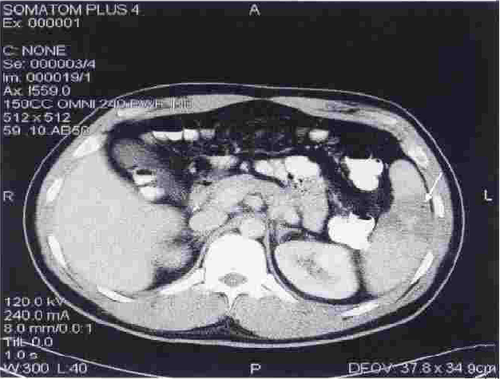
A common issue that occurs is infarction of portions of the spleen. This is actually the desired effect, as it stops the bleeding. Most of the time, we are unaware of the changes that take place in the spleen post-procedure. But every once in a while we get a repeat CT scan days or weeks down the road and see some very interesting things.

The most common finding is a splenic infarct alone. This is an area of the spleen, sometimes wedge shaped, that does not take up contrast. This is normal. In some cases, gas bubbles are seen within the spleen parenchyma, usually within the infarcted area. In others, large areas of gas are present, and an air-fluid level may also be seen. This is definitely not normal.

Note the infarcted area at the arrow, with a tiny gas bubble visible.

Tiny bubbles are normal after this procedure, and can be ignored if the patient does not appear ill and does not have any systemic evidence of inflammation or sepsis. On the other hand, big bubbles or air-fluid levels probably indicate a developing splenic abscess, and the patient will usually appear ill and have a high WBC count. Unfortunately, the only treatment for this is splenectomy. Insertion of drainage catheters does not work and the patient will only become sicker if it is attempted.

Print Friendly, PDF & Email

The 30:60 Rule For Interventional Radiology

Interventional radiology (IR) can be a very helpful adjunct to the evaluation and management of trauma patients. I’m going to talk specifically about using it for blunt trauma today because the use in penetrating trauma can be a little more nuanced.

For blunt trauma, IR is used primarily to stop bleeding. In a smaller subset of patients, this tool is used to evaluate pulse deficits. There are two basic principles that apply in either case, and I’ve wrapped them up into a single concept: the 30:60 rule for interventional radiology. 

Of course, the second law of trauma still applies: hypotensive patients cannot leave the ED to go anywhere but the OR. Once you make sure you are not violating that one, you can start the process of going to IR.

The two portions of the rule are times: the time for the IR team to arrive to start the evaluation, and the maximum time allowed for them to succeed, hence the 30:60 numbers.

The maximum acceptable time for the patient to wait until the IR team is ready for them is typically 30 minutes. US trauma center verification requires a reasonable arrival time frame, and the vast majority of hospitals have a 30 minute expectation if the team is not already in place. This response time needs to be monitored by the trauma performance improvement program (PI) and addressed if it ever exceeds the limit.

The second number is the maximum time the radiologist is given to be successful. Like other physicians, radiologists like to do a good job and finish the work they start. If they find a particularly tortuous splenic artery to navigate, they will persist at trying to get through it in order to do a selective embolization and kill the smallest piece of spleen possible. Unfortunately, this takes time and radiation (lots). And a bleeding patient is running out of time.

The good thing is that there are surgical alternatives to most of the tasks the radiologist is working on. True, some are much more difficult surgically, like managing a shattered liver or dealing with a bleeding pelvis. In those cases, I may let the interventionalist work a little longer while I keep up with blood transfusions and monitor patient status.

Bottom line:

  • Expect a 30 minute response time from the IR team
  • Let the radiologist know they have about 60 minutes to succeed. If it looks like they can’t make that, have them go to plan B (e.g. main splenic artery embolization instead of selective)
  • Make sure an experienced trauma physician is watching the patient for decompensation and is managing fluids and blood products (no pressors!)
  • If the patient decompensates at any point, they are done in IR and must proceed to OR
Print Friendly, PDF & Email

Next TraumaMedEd Newsletter Available Soon!

The March issue of TraumaMedEd is ready to go! This issue is devoted to protocols. 

Included are protocols for:

  • Anticoagulation reversal
  • TBI screening
  • Chest tube management
  • Solid organ injury management
  • And more!

Be sure to have a good QR code reader for easy retrieval. Otherwise, warm up your fingers so you can enter URLs to download the protocols.

Subscribers will get the issue delivered Saturday. It will be available to everybody on the Tuesday blog post.

Check out back issues, and subscribe now! Get it first by clicking here!

Print Friendly, PDF & Email