Category Archives: General

Wounds: When Are They Too Old To Close?

At some point in their training, every trauma professional is taught that there is a certain period time during which a wound can be safely closed. The exact number varies, but is usually somewhere between 6 and 24 hours. After that, we are told, “bad things happen.”

Always question dogma, I say. Is this true, or is it another one of those “facts” that have been propagated through the ages? Two emergency medicine groups recently performed a meta-analysis to try to answer my question. As usual, they found that much of the published literature is not very good. Out of 418 papers in their original search, only 4 fully met their criteria (laceration repaired primarily, in the ED, with clear early vs delayed criteria.

With the exception of one study with a very limited focus, there was no correlation between wound age and infection or dehiscence after primary closure. None of the studies could reliably provide a specific time beyond which closure was destined to fail. And the use of antibiotics in some of the studies also confounded the results.

Bottom line: It is more likely that infection-prone wounds get infected, not old ones. Although leaving a wound open to heal by secondary intention usually avoids the problem, it’s a big patient dissatisfier, especially with large wounds. Since many patients don’t present to the ED until their wound is “old”, it may be reasonable to try primary closure in all but infection-prone wounds. (The meaning of that phrase is not exactly clear, but most of us know it when we see it.) 

Reference: The impact of wound age on the infection rate of simple lacerations repaired in the emergency department. Injury 43(11):1793-1798, 2012.

Best Of: IV Contrast

We use CT scanning in trauma care so much that we tend to take it (and its safety) for granted. I’ve written quite a bit about thoughtful use of radiographic studies to achieve a reasonable patient exposure to xrays. But another thing to think about is the use of IV contrast.

IV contrast is a hyperosmolar solution that contains some substance (usually an iodine compound) that is radiopaque to some degree. It has been shown to have a significant impact on short-term kidney function and in some cases can cause renal failure.

Here are some facts you need to know:

  • Contrast nephrotoxicity is defined as a 25% increase in serum creatinine, usually within the first 3 days after administration
  • There is usually normal urine output and minimal to no proteinuria
  • In most cases, renal function returns to normal after 3-4 days
  • Nephrotoxicity almost never occurs in people with normal baseline kidney function
  • Large or repeated doses given within 72 hours greatly increase risk for toxicity
  • Old age and pre-existing diabetic renal impairment also greatly increase risk

If you must give contrast to a patient who is at risk, make sure they are volume expanded (tough in trauma patients), or consider giving acetylcysteine or using isosmolar contrast (controversial, may still cause toxicity).

Bottom line: If you are considering contrast CT, try to get a history to see if the patient is at risk for nephrotoxicity. Also consider all of the studies that will be needed and try to consolidate your contrast dosing. For example, you can get CT chest/abdomen/pelvis and CT angio of the neck with one contrast bolus. Consider low dose contrast injection if the patient needs formal angiographic studies in the IR suite. Always think about the global needs of your patient and plan accordingly (and safely).

Related posts:

Reference: Contrast media and the kidney. British J Radiol 76:513-518, 2003.

Trauma Mythbusters: Bathing/Showering And Wound Care

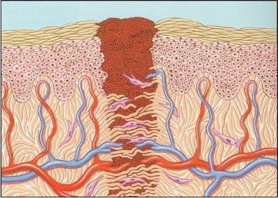
I love to hate dogma. And there’s probably nothing in surgery more sacred and more ingrained than how to take care of a wound. Everybody knows that you have to keep surgical or traumatic wounds dry, and that once you can get them wet, showers are good at baths are bad. Right?

image

And for something as common as wound management, there must be some kind of research, right? Not so! I did quite a bit of digging through the literature since 1966 and managed to find only five papers. Here are the highlights:

  • A prospective study of 100 patients were randomized to shower or bathe postoperatively. Of note, the wounds were sprayed with a clear plastic dressing before getting in the water. The was no difference in infection rates.
  • Another prospective study of 100 patients with stapled incisions after spine surgery were allowed to bathe after 2 to 5 days. Compared to historical controls, there were no differences in infection rates even though the study patients had more complex operations than controls.
  • A prospective randomized study of 121 patients after hernia surgery found no difference in infection between shower and dry groups
  • A large randomized study of 817 patients similarly showed no difference between shower and dry groups
  • Another randomized trial of 170 patients showed no difference in infections between shower after 24 hours and control groups

Get the picture? And interestingly, the few wound infections documented in any of the studies tended to occur in the dry groups, although this was not statistically significant.

Bottom line: In general, it is not harmful to get a wound wet after 24 hours. We don’t know exactly why because of the paucity of the literature, but think about it. The water that we shower or bathe in is the same water that we drink. It’s very close to sterile. When we do shower or bathe, the bacteria that come in contact with the wound are our normal skin flora, which are already in and on the wound. Plus, most incisions that have been closed are water-tight within about 24 hours. It’s more likely that using soap and water is good for you because it washes away tons of bacteria, including the pathogens!

References:
  • Prospective randomised trial of the early postoperative bathing. BMJ 19 in June 1976: 1506-1507, 1976.
  • Wound care after posterior spinal surgery. Does early grading affect the rate of wound complications? Spine (Phila PA 1976) 21(18):2160-2162, 1996.
  • Does a shower with postoperative wound healing at risk? Chirurg 68(7): 715-717, 1997.
  • Modification of postoperative wound healing by showering. Chirurg 71(2):234-236, 2000.
  • Postoperative wound healing in wound-water contact. Zentralbl Chir 125(2):157-160, 2000.

Don’t Ignore The Naughty Bits

A major part of any patient encounter is the physical exam. This is particularly true in the trauma patient, because it allows trauma professionals to identify potential life and limb threatening injuries quickly and deal with them. Unfortunately, we tend to mentally block out certain parts of the body, typically the genitalia and perineum, and may not do a complete exam of the area. I call these areas the naughty bits. For those of you who don’t get the reference, here’s the origin of this phrase:

image

Specifically, the naughty bits are the penis, vagina, perineum, anus and natal cleft (aka the butt crack or arse crack). These areas are more likely to remain covered when the patient arrives, and are less likely to be examined thoroughly.

In penetrating trauma, a detailed exam of these areas is extremely important in every patient to avoid hidden injuries and to determine if nearby internal structures (rectum, urethra) might have been injured.

Here are some tips for each of the areas:

  • Penis – Always look for any blood at the meatus (or a little blood in the underwear) as a possible sign of urethral injury. This is frequently associated with pelvic fractures.
  • Scrotum – Blood staining here is usually from blood dissecting away from pelvic fractures. Patients with this finding are more likely to need angiographic embolization of pelvic bleeding.
  • Vagina – external exam should always be done. Internal and/or speculum exam should be done in pregnant patients, and those with external bleeding or pelvic fractures
  • Perineum – Also associated with pelvic fracture and significant bleeding. Skin tears in this area are usually lacerations indicating an open pelvic fracture. Alert your orthopaedic surgeons early, and do a good, clean rectal exam (carefully wipe away all external blood). Rectal injuries are common with this finding, and a formal proctoscopic will probably be required.
  • Anus – Skin tears virtually guarantee that a deeper rectal injury will be found. Proctoscopic exam in the OR is mandatory.
  • Natal cleft – Usually not a lot going on in this area, except in penetrating injury. This is the only area of the naughty bits that can be safely examined in the lateral position. 

Bottom line: The naughty bits are important because the occasional missed injury in this area can be catastrophic! Do your job and force yourself to overcome any reluctance to examine them.

Related posts:

Pop Quiz! Final Answer

Our patient with the steak knife to the head has been evaluated by CT. The scan shows that the blade enters the right orbit, passing through the medial orbital wall into the ethmoid sinus, turbinates and nasal septum. It then passes into the left orbit along the posterior floor and exits the apex. The optic nerves are not involved, but there may be involvement of the rectus or oblique muscles to the globe. There does not appear to be any involvement of the maxillary sinus.

See why a good exam is important? Gross visual acuity and extra-ocular muscle testing is very important here. Miraculously, all are intact. So now what?

Just yank it out? Absolutely not! Although there is no gross bleeding from the nose or mouth, and none is seen on CT, that doesn’t mean there won’t be! The patient needs to go to the OR, and it may be helpful to have a facial surgeon present just in case. Scopes for evaluating the sinuses and packing materials should be readily available.

Under sedation, the knife can be smoothly withdrawn. An awake patient can tell you how it feels, and whether he is experiencing any bleeding or ocualr changes. If in doubt, the sinuses can be scoped and the globes re-examined.

Note: If troublesome bleeding does occur, this is not an area that is amenable to surgical exploration. The only realistic options available are packing and angioembolization.