Category Archives: General

Trauma Pearl: Unexpected Respiratory Failure After Blunt Trauma

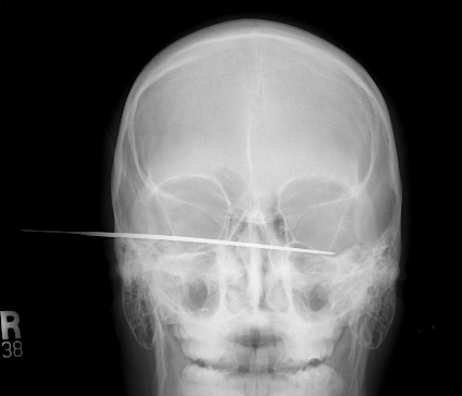
A 24 year old restrained female is involved in a T-bone type motor vehicle crash. She sustains a moderate to severe traumatic brain injury and is intubated and sedated. On exam, she has a few abrasions over her left flank, and no other physical findings. Head CT shows some subarachnoid blood, and abdominal CT is negative.

She is placed in the ICU and slowly becomes more responsive. However, her FIO2 has to be increased several times due to poor oxygenation. By day 3, she is on 90% O2 and has diffuse infiltrates in her lung fields.

What’s the problem?!

This is a classic presentation of a missed abdominal injury. Restrained patients are at risk for intestinal injuries, even with a t-bone mechanism and little to no seat belt sign. Physical exam may be helpful, but abdominal pain/tenderness may be masked by head injury.

A repeat CT scan was performed, which showed free fluid and a few bubbles of free air. The patient was taken to the OR and a bucket handle injury to the distal ileum was found, with devitalized and leaking intestine. This was resected and primary anastomosis was performed. Within 2 days, the patient was on 40% O2 and was ready for extubation two days later.

Bottom line: Unexplained respiratory failure after blunt trauma, especially if no chest injury has occurred, is nearly always due to a missed abdominal injury. The initial CT is a snapshot that is valid for only a few hours. Re-image with CT or ultrasound, and operate promptly if any significant change in patient condition occurs.


Related posts:

Fictional case, not treated at Regions Hospital.

Print Friendly, PDF & Email

Next Trauma MedEd Newsletter Available Tomorrow!

The April issue of Trauma MedEd was sent out to subscribers over the weekend. This issue, like the March issue, is devoted to protocols. 

Included are protocols for:

  • Chest tube management
  • Solid organ injury
  • Rapid reversal of warfarin
  • Reversal of other anticoagulants
  • Massive transfusion

Be sure to have a good QR code reader for easy retrieval. Otherwise, warm up your fingers so you can enter URLs to download the protocols.

This issue will be available on the blog tomorrow!

Check out back issues, and subscribe now! Get it first by clicking here!

Print Friendly, PDF & Email

How Does That Work?: Angioembolization Coils

Ever wonder how interventional radiologists stop bleeding? They are very skilled in getting access to complicated areas of the arterial tree. Once they have located a bleeding point, they’ve got to plug it up with something.

Over the years, a wide variety of things have been used. They include blood clot, tiny metal or plastic spheres, superglue, and a variety of other creative things. One of the more recent additions is the metal coil.

On xray, these look like little pieces of piano wire in various shapes after they are inserted. But how do they work? They’re metal, and fairly smooth. How does that promote fast clotting?

The answer is more obvious when you look at one of these before it’s been inserted. Note the “fuzz”. These are synthetic fibers that are wrapped into the coil itself, and they are what actually promote clotting when the coil is in place.

Print Friendly, PDF & Email

Why Do They Call It: Extra-axial Blood?

You’ve seen it on head CT reports. “The patient has a collection of extra-axial blood…” Then it goes on to describe the location and size of a subdural hematoma. But why is it called “extra-axial?”

The answer lies in the embryology of the central nervous system. Yes, it’s been a long time since any of us have read anything about that. Early animals had a straight neural tube, which slowly evolved into a brain and spinal cord. This is known as the axis of the nervous system.

The brains of early vertebrates developed at the end of the neural tube, and were oriented in the same longitudinal axis as the rest of it. As brains got bigger, a 90 degree bend occurred at the cephalic flexure.

So in humans, there is a difference between the body axis and the brain axis. But the brain axis is what really counts. This means that any blood outside of the brain axis is defined as extra-axial.

Bottom line: Extra-axial blood is defined as any bleeding outside of the brain parenchyma. This includes subdural and epidural hematomas, and subarachnoid hemorrhage. It does not include any intraparenchymal bleeding like contusions or hematomas.

Print Friendly, PDF & Email

Lab Values From Intraosseous Blood

The intraosseous access device (IO) has been a lifesaver by providing vascular access in patients who are difficult IV sticks. In some cases, it is even difficult to draw blood in these patients by a direct venipuncture. So is it okay to send IO blood to the lab for analysis during a trauma resuscitation?

A study using 10 volunteers was published last year (imagine volunteering to have an IO needle placed)! All IO devices were inserted in the proximal humerus. Here is a summary of the results comparing IO and IV blood:

  • Hemoglobin / hematocrit – good correlation
  • White blood cell count – no correlation
  • Platelet count – no correlation
  • Sodium – no correlation but within 5% of IV value
  • Potassium – no correlation
  • Choloride – good correlation
  • Serum CO2 – no correlation
  • Calcium – no correlation but within 10% of IV value
  • Glucose – good correlation
  • BUN / Creatinine – good correlation

Bottom line: Intraosseous blood can be used if blood from arterial or venous puncture is not available. Discarding the first 2cc of marrow aspirated improves the accuracy of the lab results obtained. The important tests (hemoglobin/hematocrit, glucose) are reasonably accurate, as are Na, Cl, BUN, and creatinine. The use of IO blood for type and cross is not yet widely accepted by blood banks, but can be used until other blood is available.

Related post:

Reference: A new study of intraosseous blood for laboratory analysis. Arch Path Lab Med 134(9):1253-1260, 2010.

Print Friendly, PDF & Email