Category Archives: Device

Air Embolism From an Intraosseous (IO) Line

Intraosseous (IO) lines are a godsend when we are faced with a patient who desperately needs access but has no veins. The tibia is generally easy to locate and the landmarks for insertion are straightforward. They are so easy to insert and use, we sometimes “set it and forget it”, in the words of infomercial guru Ron Popeil.

But complications are possible. The most common is an insertion “miss”, where the fluid then infuses into the knee joint or soft tissues of the leg. Problems can also arise when the tibia is fractured, leading to leakage into the soft tissues. Infection is extremely rare.

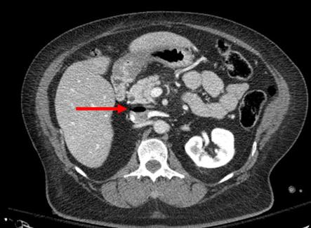
This photo shows the inferior vena cava of a patient with bilateral IO line insertions (black bubble at the top of the round IVC).

During transport, one line was inadvertently disconnected and probably entrained some air. There was no adverse clinical effect, but if the problem is not recognized and the line is not closed properly, there could be.

Bottom line: Treat an IO line as carefully as you would a regular IV. You can give anything through it that can be given via a regular IV: crystalloid, blood, drugs. And even air, so be careful!

4 Good Lab Values From Intraosseous Blood – And Some Not So Good Ones Too

The intraosseous access device (IO) has been a lifesaver by providing vascular access in patients who are difficult IV sticks. In some cases, it is even difficult to draw blood in these patients by a direct venipuncture. So is it okay to send IO blood to the lab for analysis during a trauma resuscitation?

A study using 10 volunteers was published last year (imagine volunteering to have an IO needle placed)! All IO devices were inserted in the proximal humerus. Here is a summary of the results comparing IO and IV blood:

  • Hemoglobin / hematocrit – good correlation
  • White blood cell count – no correlation
  • Platelet count – no correlation
  • Sodium – no correlation but within 5% of IV value
  • Potassium – no correlation
  • Choloride – good correlation
  • Serum CO2 – no correlation
  • Calcium – no correlation but within 10% of IV value
  • Glucose – good correlation
  • BUN / Creatinine – good correlation

Bottom line: Intraosseous blood can be used if blood from arterial or venous puncture is not available. Discarding the first 2cc of marrow aspirated improves the accuracy of the lab results obtained. The important tests (hemoglobin/hematocrit, glucose) are reasonably accurate, as are Na, Cl, BUN, and creatinine. The use of IO blood for type and cross is not yet widely accepted by blood banks, but can be used until other blood is available. NOTE: your lab may try to refuse the specimen due to “other stuff” (marrow) in the specimen. Have them run it anyway!

Reference: A new study of intraosseous blood for laboratory analysis. Arch Path Lab Med 134(9):1253-1260, 2010.

Finding Tough-To-See Veins – Revisited

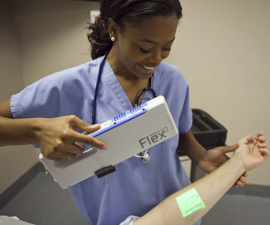
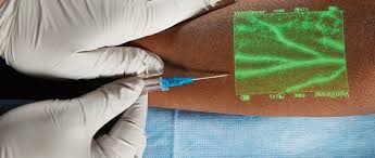
I’m always interested in technology that makes what we do easier, and this item seems to be in the news again. It’s not new technology any more; I first wrote about this way back in 2011. Here’s an objective look at an interesting machine that’s been around for a while. It uses near-infrared light to detect skin temperature changes to allow it to map out veins. It then projects an image of the map in real time onto the skin. In theory, this should make IV starts easier (as long as you can keep your head out of the way of the projector).

One of the first published papers was from Providence, Rhode Island.  It evaluated this device to see if it could simplify IV starts in a tertiary pediatric ED. It was a prospective, randomized sample of 323 children from age 0 to 17 looking at time to IV placement, number of attempts, and pain scores.

Unfortunately, the authors did not find any differences. They found that nearly 80% of IVs were started on the first attempt with or without the VeinViewer, which is less than the literature reported 2-3 attempts. This is most likely due to the level of experience of the nurses in this pediatric ED.

The authors did a planned subgroup analysis of the youngest patients (age 0-2) and found a modest decrease in IV start time (46 seconds) and the nurse’s perception of the child’s pain. Interestingly, the parents did not appreciate a difference in pain between the two groups. This may be due to the VeinViewer’s pretty green display acting as distraction therapy for the child.

The Children’s Hospital of Colorado repeated this study and reported their results earlier this year. And unfortunately they had similar findings. There were no significant differences in success rates using the VeinViewer. Also, nurses did not note any difference in their perceived insertion skills or confidence.

Bottom line: Once again, it seemed like a good idea. But that doesn’t necessarily mean that it is. And we always automatically reach for the new shiny toy. This paper points out the importance of carefully reviewing all new (read: expensive at about $20,000 each) technology before blindly implementing it. In this case, an expensive peice of equipment can’t improve upon what an experienced ED or pediatric nurse can already accomplish.
 

References: 

  1. VeinViewer-assisted intravenous catheter placement in a pediatric emergency department. Acad Emerg Med, 18(9):966-971, 2011.
  2. Utilization of a biomedical device (VeinViewer® ) to assist with peripheral intravenous catheter (PIV) insertion for pediatric nurses. J Spec Pediatr Nurs. 23(2):e12208, 2018.

I have no financial interest in Christie Digital Systems, distributor of the VeinViewer Vision®.
Off-Label Foley Use In Trauma – Part 2

Yesterday, I wrote about an unusual way to use the Foley urinary catheter to plug a heart wound. This allows you to buy time to get to the operating room to perform the definitive repair. But this cheap and effective tool is very versatile, and can be used in other body areas as well.

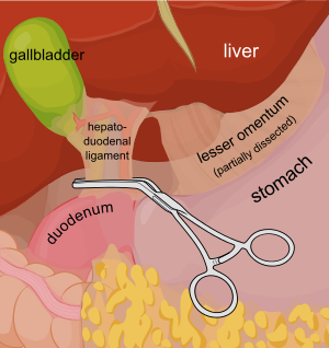
Consider a deep penetrating injury to the liver. It takes time to determine which method for slowing/stopping the bleeding is most appropriate. Sure, the doctor books say to occlude the inflow by gently clamping the hepatoduodenal ligament (Pringle maneuver). But this takes time, and can be difficult if there is lots of bleeding.

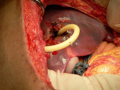
You may be able to gain some time by placing a properly sized Foley catheter directly into the wound and carefully inflating with saline. You must inflate the balloon to feel, not to its full volume. It should be snug, but not so full that it cracks the liver parenchyma and causes yet more bleeding.

Bottom line: Any time you find yourself facing bleeding from hard to expose places, think about using a balloon catheter like the Foley. Sizing is critical, and the balloon volume is more important than the catheter diameter. Estimate the size of the area that needs to be occluded, and then ask for a catheter with a 10cc or 30cc balloon. If you need smaller, more precise control, try a Fogarty arterial embolectomy catheter instead. 

As with the cardiac Foley, be sure to occlude the end so you don’t create a conduit for the blood to escape. If your patient does well, and you need to leave the catheter in place for a damage control closure, LEAVE THE CATHETER COMPLETELY WITHIN THE ABDOMEN. If you exteriorize the end, some well-meaning person may unclamp it, drop the balloon, or decide that it can be used for tube feedings.

TIP: If the distance between the balloon and the catheter tip is too long, DO NOT TRY TO SHORTEN THE TIP BY CUTTING IT! This will damage the balloon and it will not inflate.

Fogarty catheters

Air Embolism From an Intraosseous (IO) Line

Intraosseous (IO) lines are a godsend when we are faced with a patient who desperately needs access but has no veins. The tibia is generally easy to locate and the landmarks for insertion are straightforward. They are so easy to insert and use, we sometimes “set it and forget it”, in the words of infomercial guru Ron Popeil.

But complications are possible. The most common is an insertion “miss”, where the fluid then infuses into the knee joint or soft tissues of the leg. Problems can also arise when the tibia is fractured, leading to leakage into the soft tissues. Infection is extremely rare.

This photo shows the inferior vena cava of a patient with bilateral IO line insertions (black bubble at the top of the round IVC).

During transport, one line was inadvertently disconnected and probably entrained some air. There was no adverse clinical effect, but if the problem is not recognized and the line is not closed properly, there could be.

Bottom line: Treat an IO line as carefully as you would a regular IV. You can give anything through it that can be given via a regular IV: crystalloid, blood, drugs. And even air, so be careful!