Category Archives: Device

REBOA Size: Where Did The French System For Catheter Size Come From?

Medicine sure has some weird measurement systems. Besides the more standardized units like microliters, milligrams, and International Units, we’ve got some odd stuff like French (tubes) and gauge (needles). When dealing with tubes and catheters, the size is usually specified in French units.

Since I’m posting several articles on the size of REBOA (resuscitative endovascular balloon occlusion of the aorta) this week, I figured I would re-post this article on where the French sizing system came from.

Where did this crazy French system come from? It was introduced by a Swiss-born gentleman named Joseph-Frédéric-Benoît Charrière. He moved to Paris and was apprenticed to a knife maker. At the age of 17, he founded a  company that manufactured surgical instruments. His company developed and improved a number of surgical instruments, including hypodermic needles and various catheters.

Charrière introduced the system for describing catheters based on their outer diameter (OD).  It was actually named after him, and in France one will occasionally see catheters described in Ch units. Unfortunately, we Americans had a hard time pronouncing his name, and changed it to the French system (Fr).

So what’s the translation? The Ch or Fr number is the outer diameter of a catheter in millimeters multiplied by 3. It is not the outer circumference in millimeters, and the use of pi is not involved. So a big chest tube (36 Fr) has an OD of 12 mm, and a bigger chest tube (40 Fr) has an OD of 13.33 mm.

Print Friendly, PDF & Email

The Shrinking REBOA Catheter

REBOA (resuscitative endovascular balloon occlusion of the aorta) is one of the relatively new toys in our trauma toy chest. Although it’s been used for decades by vascular surgeons, believe it or not it only made the jump into the trauma world less than 10 years ago.

The original catheters used during the early days, and primarily in swine models, required a 15 French sheath for insertion. As might be expected, insertion of these huge sheaths into the common femoral artery can cause significant vascular injury. Equipment manufacturers have been steadily reducing the size of the REBOA catheter, first to 12 Fr and then to the 7 Fr size commonly used today.

The surgery group at the London Health Sciences Center in London, Ontario, Canada performed a pilot study of a new and much smaller sheath and REBOA catheter. It is made by Front Line Medical Technologies, also located in London. This was a proof of concept for the device and was performed in seven neurological death organ donors prior to their donation.

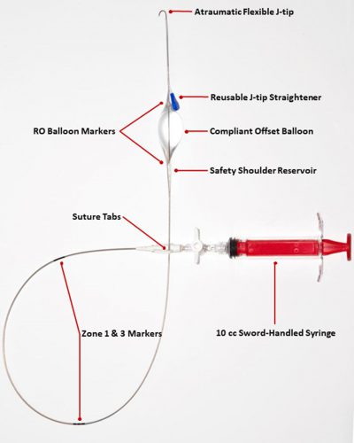
The kit consists of a 4 Fr sheath introducer with a 21 gauge needle and a guidewire, plus the REBOA catheter itself. Here is an image of the catheter:

This catheter includes several innovations not found in current catheters used in the US. I will do a side by side review of these later this week.

Here are the factoids:

  • Seven organ donors were studied after appropriate consent from the hospital IRB, organ procurement agency, organ donor procurement team, and family
  • A single general/vascular surgeon performed all insertions
  • A left sided arterial line using the 4 Fr sheath was inserted for monitoring before the procurement began
  • A right sided 4 Fr sheath was inserted for catheter insertion after the procurement incision was made
  • Average sheath insertion time was 48 seconds, and deployment time for the catheter was an average of 70 seconds (max time was 105 seconds)
  • Occlusion was confirmed by the left femoral arterial pressure monitor and by palpating the aorta below the baloon

Bottom line: This was a very simple study of the feasibility of using a smaller REBOA catheter. It measured both ease of insertion and presence of full occlusion. This is an exciting study, because there is the potential for easier insertion and fewer vascular complications at the insertion site. Obviously, these factors are not yet known, and only further work will make this clear. 

Nonetheless, easier and safer insertion has the potential to increase the use of REBOA. This will allow us to get quicker answers to the nagging questions about whether it is actually a valuable resuscitation tool and help us figure out how and in whom it is best used.

Reference: Size matters: first-in-human study of a novel 4 French REBOA device. Trauma Surgery & Acute Care Open 2021;6:e000617. doi: 10.1136/tsaco-2020-000617

Print Friendly, PDF & Email

How To Make TEG / ROTEM Useful

A lot of papers have been written on the use of thromboelastography in trauma. And pretty much any meeting or course you may attend has at least one talk on it. And I get it. It can be an important tool in treating trauma patients who have some sort of coagulation disturbance. It helps us figure out what specific part of the coagulation process is out of whack and suggests how we can fix it.

But there are a few problems. And the “friction” that those issues cause overall decreases how useful it is. Here’s a partial list of the problems:

  • The equipment costs money, and the disposables that must be used for every patient do, too.
  • Where do you put the machine? Most hospitals can’t put one unit in every possible area it might be used.
  • How to you get the results to a care area if there is no unit there?
  • There is a significant learning curve for interpreting the results
  • How can it be integrated into the massive transfusion protocol?

The main issue is that the current state of TEG and ROTEM are very similar to the state of electrocardiography shortly after it’s discovery. Here’s what you got then:

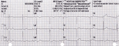
In order to get the most from an EKG, you need to combine this tracing with that from other leads, do a bunch of measurements, look for abnormal shapes and elevations/depressions, etc.  This is exactly where we are with TEG and ROTEM today. Relatively crude, and it takes a lot of work to use it.

The tracing below shows where we are with EKGs today. A computer program looks at all the tracings, and rapidly applies a complex set of rules to come to a set of diagnoses. Notice in the image below that this reading is “unconfirmed.” But how many times in your career have you seen a cardiologist correct one of these? The machines are actually very good!

Bottom line: The tracing above is where we need to be with TEG and ROTEM. Instead of a clinician staring at a developing tracing and figuring out what products to give, these machines need to be just like an automated EKG machine. Sure, a human can still stare at the trace. But the machine will automatically monitor it, apply rules about what abnormalities are present and what is needed to correct them. Send off your blood specimen, and within minutes instructions like “infuse 2 units of plasma now” or “give 12u cryo now” appear. These may be displayed on a monitor in the treatment area, or be broadcast to the phone or pager of the responsible clinicians.

Current TEG/ROTEM equipment is what I would consider 1st generation. The next generation will reduce or remove much of the “friction” in the current process and allow us to really integrate TEG/ROTEM meaningfully into the massive transfusion protocol for trauma. And for anyone who develops this 2nd generation equipment, don’t forget my royalty checks for this idea! 

In my next post, I’ll review the new EAST guidelines for the use of TEG and ROTEM.

Print Friendly, PDF & Email

Should I Apply Compression Devices To Patients With DVT?

Everyone knows that venous thromboembolism (VTE) is a potential problem in hospitalized patients, and especially so in trauma patients. Several groups of them are at higher risk by virtue of the particular injuries they have sustained and the activity restriction caused.

Nearly every trauma program uses some form of screening and prophylaxis in an attempt to reduce the occurrence of this problem, which can result in deep venous thrombosis (DVT) and/or pulmonary embolism (PE). Screening looks at patient factors such as age, obesity, previous VTE as well as injury risk factors like spine and pelvic fractures, and decreased mobility.

Based on the screening protocol, prophylaxis may be prescribed depending upon level of VTE risk, which is then balanced with bleeding risk from brain, solid organ, or other injuries. The choices we have are primarily mechanical vs chemical and consist of compression devices (sequential or not) and various heparins.

An age old question surfaced on my own patient rounds recently. If a patient breaks through their prophylaxis and develops DVT, is it safe to apply compression devices to the extremity?

There has always been the fear that doing things that increase flow in the affected extremity may cause clots to dislodge and ultimately cause a PE. Seems logical right? But we know that often, our common sense about things is completely wrong.  Couldn’t just moving around cause pieces to break off? A meta-analysis of 13 studies published in 2015 showed that early ambulation was not associated with a higher incidence of new PE. Furthermore, patients who suffered from pain in the affected extremity noted significant improvements with early ambulation.

If ambulation makes the pain better, could the veins be recanalizing more quickly? Another study examined a small group of 72 people with DVT receiving anticoagulants, half of whom were prescribed exercise and compression stockings and the other half stockings only. There was a huge amount of variability in the rates of recanalization, but ultimately there were no significant differences with or without exercise.

So just lying in bed is not good, and exercise/ambulation may actually make people feel better. But interestingly, bedrest alone does not appear to increase the likelihood of PE! It does decrease the risk of developing problems other than the VTE, like pulmonary complications.

But what about compression devices? Common sense would say that you are intermittently  increasing pressures in the leg veins, which could dislodge any loose clots and send them flying to the lungs, right?

Unfortunately, I couldn’t find a paper from anyone who had the courage to try this. Or perhaps no institutional review board (IRB) would approve it. But the key fact is that every compression device manufacturer includes existing DVT as a contraindication in their product documentation. They don’t have any literature either, so I assume it’s an attempt to limit litigation, just in case.

Bottom line: Walking provides at least as much muscle compression as compression devices. But the simple truth is that we have no solid research that either supports or condemns the use of active compression devices in patients with known DVT. And we probably won’t, ever.

Compression stockings seem to be safe, but they really don’t do much. They are white, but don’t do much more than contribute to hospital clothing fashion. Since the manufacturers define existing DVT as a contraindication, application of their product would be considered an off-label use. So it looks like we cannot in good faith use these devices in patients with diagnosed DVT.

References:

  • Bed Rest versus Early Ambulation with Standard Anticoagulation in The Management of Deep Vein Thrombosis: A Meta-Analysis. PLOS One , April 10, 2015, https://doi.org/10.1371/journal.pone.0121388
  • Bed Rest or Ambulation in the Initial Treatment of Patients With Acute Deep Vein Thrombosis or Pulmonary Embolism: Findings From the RIETE Registry. Chest 127(5):1631-1636, 2005.
  • Does supervised exercise after deep venous thrombosis improve recanalization of occluded vein segments? A randomized study. J Thrombosis Thrombolysis 23:25-30, 2006.
Print Friendly, PDF & Email

The “Double-Barrel” IO: Can It Work?

Intraosseous lines (IO) make life easy. They are quicker to insert, have a higher success rate, and require less experience than a standard IV. And they can be used for pretty much any solution or drug that can be given through an IV.

But there are some limitations. They can’t be inserted into a fractured bone. The manufacturer cautions against multiple insertions into the same bone. A second insertion should not be performed in the same bone within 48 hours.

But, as with so many things in medicine, there is little in the way of proof for these assertions. They seem like good ideas for precautions, but that does not mean they are correct. No real research has been done in this area. Until now.

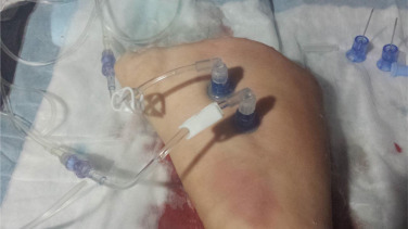
The concept of using two IO needles in one bone was explored in an animal model by researchers in Canada. They used a swine model (using the foreleg/humerus, to be exact), and tested several infusion setups.

Here are the factoids:

  • Infusing crystalloid using an infusion pump set to 999ml/hr took 30 minutes with a single IO, and 15 minutes with a “double-barrel” setup
  • Giving crystalloid using a pressure bag set at 300 mm/Hg took 24 minutes with a single IO, and 23 minutes with double the fun
  • The double-barrel setup also worked for a blood/drug combo. 250cc of blood and 1 gm of TXA in 100ml of saline infused via pump in 13 minutes.
  • Simultaneous anesthesia drugs (ketamine infusion in IO #1, fentanyl and rocuronium bolus in IO #2) without problems
  • Multiple fluid + drug infusion combinations were tested without incident
  • There were no needle dislodgements, soft tissue injuries, fractures, or macrohistologic damage to the bone or periosteum

Bottom line: Remember, these are pigs. Don’t do this in humans yet. However, this is pretty compelling evidence that the double-barrel IO concept will work in people. And it appears that infusion pumps must be used for effective, fast infusions. I recommend that prehospital agencies with inquiring minds set up a study in people to prove that this works in us, too.

Reference: Double-barrelled resuscitation: A feasibility and simulation study of dual-intraosseous needles into a single humerus. Injury 46(11):2239-42, 2015.

Print Friendly, PDF & Email