Category Archives: Abdomen

Video: Minimally Invasive Repair Of Rectal Injuries

Extraperitoneal rectal injury repair has evolved considerably over the past 40 years. Way back when, this injury automatically triggered exploration, diverting colostomy with washout of the distal colon, and presacral drain insertion (remember those?).

We eventually backed off on the presacral drains (pun intended), which didn’t make a lot of sense anyway. And we gave up on dissecting down deep into the pelvis to approach the injury. This only served to contaminate an otherwise pristine peritoneal cavity. Ditto for the distal rectal washout. So we have been performing a diverting colostomy as the primary method of treatment for years.

A Brief Report in the British Medical Journal Open shows us what may very well be the next stage in treating these injuries. Whereas they were previously left to heal on their own followed by colostomy closure after a few months, these authors from Sunnybrook Health Sciences Centre in Toronto are promoting a minimally invasive approach to definitive management.

They detail two cases, one an impalement by a steel rod through the rectum and bladder, and one stab to the buttock. The authors dealt with the non-rectal injuries using conventional techniques. The rectal injuries were repaired using trans-anal minimally invasive surgery (TAMIS). Both were discharged without complications.

Here is a link to the video of the technique used in the stab victim:

Click here for video

Bottom line: It’s about time! As long as there is not a destructive injury to the extraperitoneal rectum, this seems like a great technique to try. It may very well eliminate the need for a diverting colostomy.

But remember, this is only a case report. We don’t know about antibiotic duration, followup imaging, longer term complications, or anything really. A larger series of cases is warranted to provide these answers. This will take some time due to the low frequency of this injury. So if you try it, build your own series and publish it so we all can learn!

Reference: Minimally invasive approach to low-velocity penetrating  extraperitoneal rectal trauma. BMJ Open 5(1) epub 5/12/2020.

Activity Guidelines After Solid Organ Injury: How Important Are They?

Just about every practice guideline out there regarding liver and spleen injury has some type of physical activity guidelines associated with it. The accepted dogma is that moving around too much, or climbing stairs, or lifting objects, or getting tackled while playing rugby could exacerbate the injury and lead to complications or surgery.

But is it true? Activity restrictions after solid organ injury have been around longer than I have been a trauma surgeon. And the more people I poll on what they do, the more and very different answers I get. And there are no decent papers published that look critically at this question. Until now. 

A pediatric multi-center study of study on adherence to activity restrictions was published last year. Ten Level I pediatric trauma centers in the US tabulated their experience with solid organ injuries over a 3.75 year period fro 2013 to 2016. Only patients with successful nonoperative management of their injury were included, and those with high grade renal or pancreatic injuries were excluded.

Since this was a pediatric study, the American Pediatric Surgical Association (APSA) practice guideline was followed (activity restriction = organ injury grade + 2 weeks). Activity restrictions included all sports, any recreational activity with wheels, or any activity that involved having both feet off the ground. Patients with Grade III-V injuries were seen at an office visit after 2 weeks, and lower grade injuries had a phone followup.

Adherence to guidelines was assessed by a followup phone call two months after injury. Clinical outcomes assessed at 60 days included unplanned return to the emergency department (ED), re-admission, complications, and development of new bleeding confirmed by surgery, ultrasound, or computed tomography (CT) at 60 days post injury.

Here are the factoids:

  • Of the 1007 patients in the study, some 56% were either excluded (178) or lost to followup (463)
  • Of the remaining 366, roughly 46% had a liver injury, 44% spleen, and the remaining 10% had both
  • Median age was 10, so this was actually a younger population
  • 76% of patients claimed they abided by the guidelines, 14% said they did not, and 10% “didn’t know.” This means they probably did not.
  • For the 279 patients who said they adhered to activity restrictions 13% returned to the ED and half were admitted to the hospital
  • Of the 49 patients who admitted they did not follow the guidelines, 8% returned to the ED at some point and none were readmitted
  • The most common reasons for return to ED were abdominal pain, anorexia, fatigue, dizziness, and shoulder pain
  • There were no delayed operations in either of the groups

Bottom line: There were no significant differences between the compliant and noncompliant groups. Unfortunately, the authors did not include an analysis of the “I don’t know if I complied” group, which would have been interesting. But there is one issue I always worry about in these low-number-of-subjects studies that don’t show a significant difference between groups. Did they have the statistical power to show such a difference? If not, then we still don’t know the answer. And unfortunately, I’m not able to guess the numbers well enough to do the power calculation for this study.

I am still intrigued by this study! Our group originally had a fixed time period (6 weeks) of limited activity in our practice guideline for pediatric solid organ injury patients. This was rescinded last year based on our experience of no delayed complications and guidance from our sister pediatric trauma center at Children’s Hospital in Minneapolis. We are also moving toward making a similar change on our adult practice guideline.

Too many centers wait too long to make changes in their practice guidelines. They bide their time waiting for new, published research that they can lean on for their changes. Unfortunately, I think they will be waiting for a long time because many of our questions are not interesting enough for acceptance by the usual journals. Rely on the expertise and experience of your colleagues and then make those changes. Be sure to follow with your performance improvement program to make sure that they actually do work as well as you think!

Reference: Adherence to APSA activity restriction guidelines and 60-day clinical outcomes for pediatric blunt liver and splenic injuries (BLSI). J Ped Surg 54:335-339, 2019.

4 Guidelines For the Management Of Bladder Injury

The Eastern Association for the Surgery of Trauma (EAST) has been at the forefront of trauma practice guideline dissemination for decades. They recently published a set of recommendations for managing patients with bladder injury. These injuries are not commonly encountered by trauma professionals, and I thought a refresher on current thinking on their management was in order.

Using the usual methodology, the trauma literature was scanned for papers dealing with this topic. After screening for quality, the field was narrowed to 17 papers which were used to formulate the published recommendations. These cover imaging and management questions that frequently come up during the evaluation of these patients.

Following are the questions raised, the EAST recommendations, and my commentary about them:

    • In patients with abdominal / pelvic trauma, should retrograde CT cystography vs no imaging be used to diagnose a bladder injury? This seems like a silly question, but the answer lies in the details. It all boils down to the likelihood of injury. And how does one determine likelihood? By looking at the urine and the fracture patterns around the bladder. Patients with microscopic hematuria are very unlikely to have a bladder injury, and any type of bladder imaging in these patients (cystogram, CT cystogram) is almost never positive, and so is not indicated. This is the reason that ordering a urinalysis in major trauma patients is not recommended. However, if gross hematuria is present, CT cystogram is recommended. The sensitivity and specificity are nearly perfect. Just be sure to do a true cystogram by actively filling the bladder with contrast via a urinary catheter. Passive filling of the bladder with urine from the IV contrast misses about half of all the injuries. Also, strongly consider adding CT cystogram in patients with widening of the pubic symphysis. This injury pattern is frequently associated with bladder injury.
    • In patients with intraperitoneal bladder rupture from blunt trauma, should operative or nonoperative management be used to decrease complications? Another silly question? In general, intraperitoneal bladder ruptures do not heal on their own, so urine continues to bathe the peritoneal cavity until the injury is fixed. The review article recommended that operative repair be performed in all of these cases. 
    • In patients with extraperitoneal bladder rupture from blunt trauma, should operative or nonoperative management be used to decrease complications? Patients with a simple extraperitoneal bladder injury should undergo nonoperative management. These injuries usually heal and seal within about 10 days. However, patients with this type of bladder injury that is more complicated (bone spicules piercing the bladder, concomitant vaginal or rectal injury, bladder neck injury) should undergo operative repair in order to decrease the complication rate. One additional group that should be repaired: patients with pubic diastasis that will require operative fixation. The bladder should be repaired at the time of the orthopedic procedure to avoid bathing the new hardware in urine.
    • In patients who have undergone operative or nonoperative management of bladder injury, should bladder closure be assessed with cystogram or not? This one depends on the type and complexity of injury. For simple intraperitoneal bladder injuries that were operatively repaired, no followup cystogram is required. More complex repairs should be evaluated by cystogram before removing the urinary catheter. Finally, simple extraperitoneal injuries should also have a cystogram obtained before removing the catheter. My magic number for obtaining followup studies is 10 days. There is no real science behind this, and no one has systematically looked at 5 vs 7 vs 10 vs 14 days. This one is based only on personal experience.

And by the way, most simple bladder injuries (both intra- and extra-peritoneal) can be easily repaired using two layers by your friendly neighborhood trauma surgeon. More complex injuries are generally best left to the urologist.

Reference: Management of blunt force bladder injuries: A practice management guideline from the Eastern Association for the Surgery of Trauma. J Trauma 86(2):326-336, 2019.

Spleen Vaccines: So Confusing! – Part 2

Who needs to get these vaccines? Obviously if your patient’s spleen was surgically removed, they should get it. But what about patients who underwent angioembolization? Unfortunately, the only data available is either very old or is based on antibody response to the vaccine. And antibody titers do not predict immunity to infection, so these studies are close to meaningless.

Old research showed that the spleen’s immune function was preserved as long as 50% of its blood flow was delivered through the splenic artery. How can you tell if half of the spleen is still functioning after splenic angioembolization? Look at the images and make an educated guess. If in doubt, vaccinate.

When is the best time to vaccinate? There has been much gnashing of teeth regarding early vs late vaccination. The arguments against early vaccination center around the typical immune suppression seen with major trauma. However, trauma patients frequently do not appear for all their followup visits and would not receive vaccines at all if they are a no-show. So I recommend vaccinating as early as possible during the hospital stay to avoid forgetting. The data recommending waiting until just before discharge are also based on antibody titers, and I don’t buy it.

Bottom line: I’m not an epidemiologist. But making a set of vaccination rules more complicated for a complex population seems unwise. Especially since the added vaccine offers protection for only one more serotype of Pneumococcus.

But I can’t argue with the FDA and CDC. I have no idea of the wheeling and dealing that occurred to get the new vaccine approved. All we can do is follow the recommendations the best we can, and try to remind our patients to get that Pneumovax and meningococcal conjugate booster five years down the road. Good luck with that.

Spleen Vaccines: So Confusing! – Part 1

Earlier this year, there were a lot of television commercials for Prevnar 13, a 13-valent pneumococcal vaccine for immun-ocompromised or asplenic adults. And interestingly, I noticed that the CDC has added a recommendation that these patients receive this vaccination, followed by the original 23-valent vaccine (Pneumovax 23) 8 weeks later.

WTF? Patients with splenectomy (or significant angio-embolization) for trauma are considered functionally asplenic. And although the data for immunization in this group is weak, giving triple vaccinations with pneumcoccal, H. flu, and meningococcal vaccines has become a standard of care.

This was difficult enough already because there was debate around the best time to administer: during the hospital stay or several weeks later after the immune system depression from trauma had resolved. The unfortunate truth is that many trauma patients never come back for followup, and so don’t get any vaccines if they are not given during the hospital stay.

And then came the recommendation a few years ago to give a 5-year booster for the pneumococcal vaccine. I have a hard time remembering when my last tetanus vaccine was to schedule my own booster. How can I expect my trauma patients to remember and come back for their pneumococcal vaccine booster?

So what do we do with the CDC Prevnar 13 recommendation? If we add it, it means that we give Prevnar while the patient is in the hospital, and then hope they come back 8 weeks later for their Pneumovax. And then 5 years later for the booster dose. Huh?

Looking at the package insert, I read that Pneumovax 23 protects against 23 serotypes of S. Pneumo, which represent 85% of most commonly encountered strains out there. So it’s not perfect. Prevnar 13 protects against 13 serotypes, and there is no in-dication as to what percent of strains encountered are protected against.

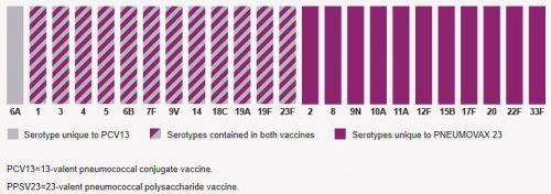
So I decided to dig deeper and look at the serotypes included in each vaccine. They are shown in the chart below. The 23 bars with maroon in them (solid or striped) are Pneumococcal serotypes covered by Pneumovax 23. The 13 bars containing gray are ones covered by Prevnar 13. There is only one serotype in Prevnar 13 not covered by Pneumovax 23, serotype 6A. Unfortunately, it’s nearly impossible to find the prevalence of infections by serotype, and it varies geographically and over time anyway. So does cover-age of a single extra serotype by Prevnar 13 justify an additional vaccination and complicated administration schedule? Hmm.

It turns out that there is one significant difference between these two vaccines. Pneumovax 23 is a polysaccharide vaccine made up of fragments of polysaccharide from pneumococcus cell walls. Prevnar 13 is a “conjugated vaccine,” meaning that the polysaccharides are linked to a protein. This is thought to increase the immune system response to the vaccine.

(click for full-size graph)

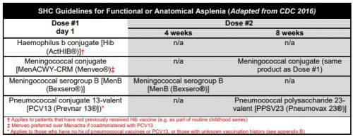
The current CDC recommendations are listed below. In the old days, we just gave three vaccines before the patient left the hospital. Then the Pneumovax 23 booster was added at 5 years. Same for the meningococcal serogroup B booster at 4 weeks. Then the meningococcal conjugate vaccine (Menactra) came along and was added (with a booster at 8 weeks). Finally, Prevnar 13 was added with its own booster, and Pneumovax 23 was delayed for 8 weeks. Oh, and don’t forget the 5 year boosters for both Pneumovax 23 and the meningococcal conjugate vaccines. It has become very complicated.

(click for full-size chart)

In my next post, I’ll try to make sense of this mish-mash and offer some thoughts on how to decide what to do for your patients.